Movie
Movie Controller

[English] 日本語
 Yorodumi
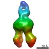
- EMDB-4536: Nanodisc reconstituted Human-mouse chimeric ABCB1 (ABCB1HM)-EQ mu... -
+ Open data

- Basic information
| Entry | Database: EMDB / ID: EMD-4536 | |||||||||
|---|---|---|---|---|---|---|---|---|---|---|
| Title | Nanodisc reconstituted Human-mouse chimeric ABCB1 (ABCB1HM)-EQ mutant in complex with UIC2 Fab and Zosuquidar. | |||||||||
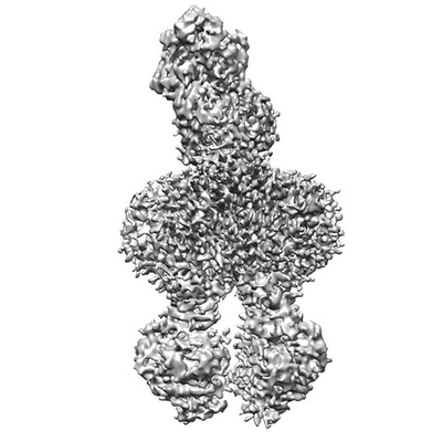
 Map data Map data | Postprocessed map of human-mouse chimeric ABCB1 (ABCB1HM)- EQ mutant in complex with UIC2 fab and zosuquidar | |||||||||
 Sample Sample |
| |||||||||
 Keywords Keywords | ABCB1 / p-glycoprotein / p-gp / multidrug transporter / ABC transporter / zosuquidar / membrane transporter / MEMBRANE PROTEIN | |||||||||
| Biological species |  Homo sapiens (human) / Homo sapiens (human) /  | |||||||||
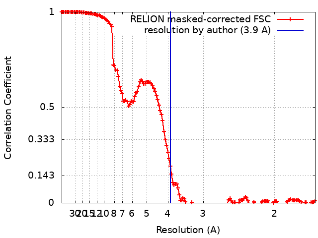
| Method | single particle reconstruction / cryo EM / Resolution: 3.9 Å | |||||||||
 Authors Authors | Alam A | |||||||||
| Funding support |  Switzerland, 2 items Switzerland, 2 items
| |||||||||
 Citation Citation | Journal: Science / Year: 2019 Title: Structural insight into substrate and inhibitor discrimination by human P-glycoprotein. Authors: Amer Alam / Julia Kowal / Eugenia Broude / Igor Roninson / Kaspar P Locher /  Abstract: ABCB1, also known as P-glycoprotein, actively extrudes xenobiotic compounds across the plasma membrane of diverse cells, which contributes to cellular drug resistance and interferes with therapeutic ...ABCB1, also known as P-glycoprotein, actively extrudes xenobiotic compounds across the plasma membrane of diverse cells, which contributes to cellular drug resistance and interferes with therapeutic drug delivery. We determined the 3.5-angstrom cryo-electron microscopy structure of substrate-bound human ABCB1 reconstituted in lipidic nanodiscs, revealing a single molecule of the chemotherapeutic compound paclitaxel (Taxol) bound in a central, occluded pocket. A second structure of inhibited, human-mouse chimeric ABCB1 revealed two molecules of zosuquidar occupying the same drug-binding pocket. Minor structural differences between substrate- and inhibitor-bound ABCB1 sites are amplified toward the nucleotide-binding domains (NBDs), revealing how the plasticity of the drug-binding site controls the dynamics of the adenosine triphosphate-hydrolyzing NBDs. Ordered cholesterol and phospholipid molecules suggest how the membrane modulates the conformational changes associated with drug binding and transport. | |||||||||
| History |
|
- Structure visualization
| Movie |
 Movie viewer Movie viewer |
|---|---|
| Structure viewer | EM map: SurfViewMolmilJmol/JSmol |
| Supplemental images |
- Downloads & links
-EMDB archive
| Map data | emd_4536.map.gz | 228.6 MB | EMDB map data format | |
|---|---|---|---|---|
| Header (meta data) | emd-4536-v30.xmlemd-4536.xml | 21.6 KB 21.6 KB | Display Display | EMDB header |
| FSC (resolution estimation) | emd_4536_fsc.xml | 14.2 KB | Display | FSC data file |
| Images |  emd_4536.png emd_4536.png | 66 KB | ||
| Filedesc metadata | emd-4536.cif.gz | 8 KB | ||
| Archive directory |  http://ftp.pdbj.org/pub/emdb/structures/EMD-4536ftp://ftp.pdbj.org/pub/emdb/structures/EMD-4536 http://ftp.pdbj.org/pub/emdb/structures/EMD-4536ftp://ftp.pdbj.org/pub/emdb/structures/EMD-4536 | HTTPS FTP |
-Related structure data
| Related structure data |  6qeeMC  4539C 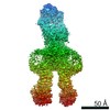 4540C 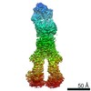 4541C  6qexC M: atomic model generated by this map C: citing same article ( |
|---|---|
| Similar structure data |


-Links
| EMDB pages | EMDB (EBI/PDBe) / EMDataResource |
|---|
-Map
| File | Download / File: emd_4536.map.gz / Format: CCP4 / Size: 244.1 MB / Type: IMAGE STORED AS FLOATING POINT NUMBER (4 BYTES) | ||||||||||||||||||||||||||||||||||||||||||||||||||||||||||||||||||||
|---|---|---|---|---|---|---|---|---|---|---|---|---|---|---|---|---|---|---|---|---|---|---|---|---|---|---|---|---|---|---|---|---|---|---|---|---|---|---|---|---|---|---|---|---|---|---|---|---|---|---|---|---|---|---|---|---|---|---|---|---|---|---|---|---|---|---|---|---|---|
| Annotation | Postprocessed map of human-mouse chimeric ABCB1 (ABCB1HM)- EQ mutant in complex with UIC2 fab and zosuquidar | ||||||||||||||||||||||||||||||||||||||||||||||||||||||||||||||||||||
| Projections & slices | Image control
Images are generated by Spider. | ||||||||||||||||||||||||||||||||||||||||||||||||||||||||||||||||||||
| Voxel size | X=Y=Z: 0.84 Å | ||||||||||||||||||||||||||||||||||||||||||||||||||||||||||||||||||||
| Density |
| ||||||||||||||||||||||||||||||||||||||||||||||||||||||||||||||||||||
| Symmetry | Space group: 1 | ||||||||||||||||||||||||||||||||||||||||||||||||||||||||||||||||||||
| Details | EMDB XML:
CCP4 map header:
| ||||||||||||||||||||||||||||||||||||||||||||||||||||||||||||||||||||
 Z (Sec.)
 Y (Row.)
 X (Col.)


-Supplemental data
- Sample components
+Entire : Nanodisc reconstituted ABCB1HM (human mouse chimeric ABCB1) EQ mu...
+Supramolecule #1: Nanodisc reconstituted ABCB1HM (human mouse chimeric ABCB1) EQ mu...
+Supramolecule #2: ABCB1HM
+Supramolecule #3: UIC2 Fab
+Macromolecule #1: ABCB1HM-EQ
+Macromolecule #2: UIC2 Antigen Binding Fragment Light chain
+Macromolecule #3: UIC2 Antigen Binding Fragment Heavy Chain
+Macromolecule #4: 2-acetamido-2-deoxy-beta-D-glucopyranose

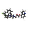
+Macromolecule #5: Zosuquidar
+Macromolecule #6: CHOLESTEROL

+Macromolecule #7: 1,2-Distearoyl-sn-glycerophosphoethanolamine

-Experimental details
-Structure determination
| Method | cryo EM |
|---|---|
 Processing Processing | single particle reconstruction |
| Aggregation state | particle |
-Sample preparation
| Concentration | 0.2 mg/mL |
|---|---|
| Buffer | pH: 7.5 |
| Grid | Model: Quantifoil R1.2/1.3 / Material: COPPER |
| Vitrification | Cryogen name: ETHANE-PROPANE / Chamber humidity: 100 % / Chamber temperature: 277 K / Instrument: FEI VITROBOT MARK IV |
- Electron microscopy
| Microscope | FEI TITAN KRIOS |
|---|---|
| Image recording | Film or detector model: GATAN K2 SUMMIT (4k x 4k) / Detector mode: SUPER-RESOLUTION / Average electron dose: 2.1 e/Å2 |
| Electron beam | Acceleration voltage: 300 kV / Electron source:  FIELD EMISSION GUN FIELD EMISSION GUN |
| Electron optics | Illumination mode: FLOOD BEAM / Imaging mode: BRIGHT FIELD |
| Experimental equipment |  Model: Titan Krios / Image courtesy: FEI Company |