Movie
Movie Controller


+ Open data

- Basic information
| Entry | Database: EMDB / ID: EMD-4504 | ||||||||||||
|---|---|---|---|---|---|---|---|---|---|---|---|---|---|
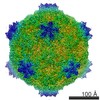
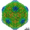
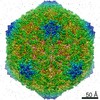
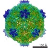
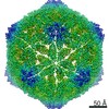
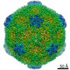
| Title | Cryo-EM Atomic Structure of Broad Bean Stain Virus (BBSV) | ||||||||||||
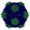
 Map data Map data | sharpened map | ||||||||||||
 Sample Sample |
| ||||||||||||
 Keywords Keywords | comovirus capsid plant virus BBSV / virus | ||||||||||||
| Function / homology |  Function and homology information Function and homology informationtransport of virus in host, cell to cell / host cell plasmodesma / T=3 icosahedral viral capsid / symbiont-mediated suppression of host innate immune response / GTP binding / structural molecule activity / DNA binding / RNA binding Similarity search - Function | ||||||||||||
| Biological species |  Broad bean stain virus Broad bean stain virus | ||||||||||||
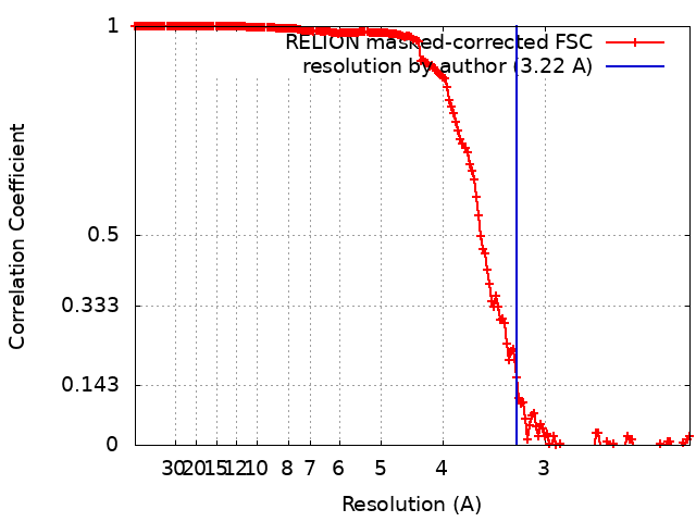
| Method | single particle reconstruction / cryo EM / Resolution: 3.22 Å | ||||||||||||
 Authors Authors | Lecorre F / Lai Jee Him J / Blanc S | ||||||||||||
| Funding support | New Caledonia,  France, 3 items France, 3 items
| ||||||||||||
 Citation Citation | Journal: Virology / Year: 2019 Title: The cryo-electron microscopy structure of Broad Bean Stain Virus suggests a common capsid assembly mechanism among comoviruses. Authors: François Lecorre / Joséphine Lai-Kee-Him / Stéphane Blanc / Jean-Louis Zeddam / Stefano Trapani / Patrick Bron / Abstract: The Broad bean stain virus (BBSV) is a member of the genus Comovirus infecting Fabaceae. The virus is transmitted through seed and by plant weevils causing severe and widespread disease worldwide. ...The Broad bean stain virus (BBSV) is a member of the genus Comovirus infecting Fabaceae. The virus is transmitted through seed and by plant weevils causing severe and widespread disease worldwide. BBSV has a bipartite, positive-sense, single-stranded RNA genome encapsidated in icosahedral particles. We present here the cryo-electron microscopy reconstruction of the BBSV and an atomic model of the capsid proteins refined at 3.22 Å resolution. We identified residues involved in RNA/capsid interactions revealing a unique RNA genome organization. Inspection of the small coat protein C-terminal domain highlights a maturation cleavage between Leu567 and Leu568 and interactions of the C-terminal stretch with neighbouring small coat proteins within the capsid pentameric turrets. These interactions previously proposed to play a key role in the assembly of the Cowpea mosaic virus suggest a common capsid assembly mechanism throughout all comovirus species. | ||||||||||||
| History |
|
- Structure visualization
| Movie |
Movie viewer |
|---|---|
| Structure viewer | EM map: SurfViewMolmilJmol/JSmol |
| Supplemental images |

- Downloads & links
-EMDB archive
| Map data | emd_4504.map.gz | 447 MB | EMDB map data format | |
|---|---|---|---|---|
| Header (meta data) | emd-4504-v30.xmlemd-4504.xml | 20.8 KB 20.8 KB | Display Display | EMDB header |
| FSC (resolution estimation) | emd_4504_fsc.xml | 17.6 KB | Display | FSC data file |
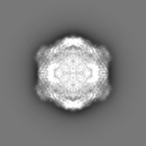
| Images | 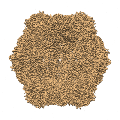 emd_4504.png emd_4504.png | 227 KB | ||
| Filedesc metadata | emd-4504.cif.gz | 6.4 KB | ||
| Others | emd_4504_additional.map.gzemd_4504_half_map_1.map.gzemd_4504_half_map_2.map.gz | 378.7 MB 380.2 MB 380.2 MB | ||
| Archive directory |  http://ftp.pdbj.org/pub/emdb/structures/EMD-4504ftp://ftp.pdbj.org/pub/emdb/structures/EMD-4504 http://ftp.pdbj.org/pub/emdb/structures/EMD-4504ftp://ftp.pdbj.org/pub/emdb/structures/EMD-4504 | HTTPS FTP |
-Related structure data
| Related structure data | 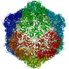 6qccMC M: atomic model generated by this map C: citing same article ( |
|---|---|
| Similar structure data |


-Links
| EMDB pages | EMDB (EBI/PDBe) / EMDataResource |
|---|---|
| Related items in Molecule of the Month |

-Map
| File | Download / File: emd_4504.map.gz / Format: CCP4 / Size: 476.8 MB / Type: IMAGE STORED AS FLOATING POINT NUMBER (4 BYTES) | ||||||||||||||||||||||||||||||||||||||||||||||||||||||||||||
|---|---|---|---|---|---|---|---|---|---|---|---|---|---|---|---|---|---|---|---|---|---|---|---|---|---|---|---|---|---|---|---|---|---|---|---|---|---|---|---|---|---|---|---|---|---|---|---|---|---|---|---|---|---|---|---|---|---|---|---|---|---|
| Annotation | sharpened map | ||||||||||||||||||||||||||||||||||||||||||||||||||||||||||||
| Projections & slices | Image control
Images are generated by Spider. | ||||||||||||||||||||||||||||||||||||||||||||||||||||||||||||
| Voxel size | X=Y=Z: 1.11 Å | ||||||||||||||||||||||||||||||||||||||||||||||||||||||||||||
| Density |
| ||||||||||||||||||||||||||||||||||||||||||||||||||||||||||||
| Symmetry | Space group: 1 | ||||||||||||||||||||||||||||||||||||||||||||||||||||||||||||
| Details | EMDB XML:
CCP4 map header:
| ||||||||||||||||||||||||||||||||||||||||||||||||||||||||||||
 Z (Sec.)
 Y (Row.)
 X (Col.)


-Supplemental data
-Additional map: unsharpened map
| File | emd_4504_additional.map | ||||||||||||
|---|---|---|---|---|---|---|---|---|---|---|---|---|---|
| Annotation | unsharpened map | ||||||||||||
| Projections & Slices |
| ||||||||||||
| Density Histograms |


-Half map: #2
| File | emd_4504_half_map_1.map | ||||||||||||
|---|---|---|---|---|---|---|---|---|---|---|---|---|---|
| Projections & Slices |
| ||||||||||||
| Density Histograms |


-Half map: #1
| File | emd_4504_half_map_2.map | ||||||||||||
|---|---|---|---|---|---|---|---|---|---|---|---|---|---|
| Projections & Slices |
| ||||||||||||
| Density Histograms |


- Sample components
-Entire : Broad bean stain virus
| Entire | Name: Broad bean stain virus |
|---|---|
| Components |
|
-Supramolecule #1: Broad bean stain virus
| Supramolecule | Name: Broad bean stain virus / type: virus / ID: 1 / Parent: 0 / Macromolecule list: all / NCBI-ID: 593572 / Sci species name: Broad bean stain virus / Virus type: VIRION / Virus isolate: STRAIN / Virus enveloped: No / Virus empty: No |
|---|---|
| Host (natural) | Organism:   Vicia faba (fava bean) Vicia faba (fava bean) |
| Virus shell | Shell ID: 1 / Name: capsid / Diameter: 300.0 Å / T number (triangulation number): 1 |
-Macromolecule #1: Large coat-protein subunit
| Macromolecule | Name: Large coat-protein subunit / type: protein_or_peptide / ID: 1 / Number of copies: 1 / Enantiomer: LEVO |
|---|---|
| Source (natural) | Organism: Broad bean stain virus |
| Molecular weight | Theoretical: 41.247977 KDa |
| Sequence | String: MDVDLFKLSL DDTSSVKGSL LDTRFAQVRV VIPKAMAGGN ELLNSNLYDI LVVDNNFRAA AALAHTHIIE GQIKCVCTIN LPENTGCCL ALCVNSSNRG QFSTDIYTIG SQDRMLWNPA CSKNSTFTFN PNPCGTGWSL EFLRRTKFHI SVVCVSGWSA Q PQTDLVMT ...String: MDVDLFKLSL DDTSSVKGSL LDTRFAQVRV VIPKAMAGGN ELLNSNLYDI LVVDNNFRAA AALAHTHIIE GQIKCVCTIN LPENTGCCL ALCVNSSNRG QFSTDIYTIG SQDRMLWNPA CSKNSTFTFN PNPCGTGWSL EFLRRTKFHI SVVCVSGWSA Q PQTDLVMT MDFFVANVPC VPRIYNLGSP GQTLWLNRWM GKLSFGQGVS NDIKSMPLAI GGGAGAKDSI LMNMTNAYLS LW RYFHGDL VFEVNKMSSP YIKSTVTFFI GFGGVSFQPE LEDFPNKLVQ FSEVQEKIEL KFTRAEFLTA WSTQVDPAAQ LAN DGCPYL YAMVHDSTAS TIVGDFNLGV TLTRIENFAG IGCNPGIQGA RLLGSAIATP Q UniProtKB: RNA2 polyprotein |
-Macromolecule #2: Small coat-protein subunit
| Macromolecule | Name: Small coat-protein subunit / type: protein_or_peptide / ID: 2 / Number of copies: 1 / Enantiomer: LEVO |
|---|---|
| Source (natural) | Organism: Broad bean stain virus |
| Molecular weight | Theoretical: 23.589662 KDa |
| Sequence | String: NAVVRSSPGI YSNCFSLRAP LKPDGPKSFT CDLMGGGVVT DGDTGWQVTV RNTPVSNLLR TAAWKRGTVH VQVVLAGASV KRSDWDSTV QIFLRQSMAT SSYDAKIWDI CQPGAAMLEF SFDVVGPNSG FEMWDSNWAS QTSWFLEFLI SNPAQNTLFE V NLRLDENF ...String: NAVVRSSPGI YSNCFSLRAP LKPDGPKSFT CDLMGGGVVT DGDTGWQVTV RNTPVSNLLR TAAWKRGTVH VQVVLAGASV KRSDWDSTV QIFLRQSMAT SSYDAKIWDI CQPGAAMLEF SFDVVGPNSG FEMWDSNWAS QTSWFLEFLI SNPAQNTLFE V NLRLDENF SVAGTTLMPP FVLDRVSVAR PLLGKQTKTV ARSARVVRET KEASESP UniProtKB: RNA2 polyprotein |
-Experimental details
-Structure determination
| Method | cryo EM |
|---|---|
 Processing Processing | single particle reconstruction |
| Aggregation state | particle |
-Sample preparation
| Concentration | 2 mg/mL |
|---|---|
| Buffer | pH: 7.4 / Component - Concentration: 20.0 mM / Component - Formula: K2HPO4/KH2PO4 / Component - Name: Phosphate |
| Vitrification | Cryogen name: ETHANE / Chamber humidity: 99 % / Chamber temperature: 298.15 K / Instrument: GATAN CRYOPLUNGE 3 / Details: blot for 1 second before plunging. |
- Electron microscopy
| Microscope | FEI TITAN KRIOS |
|---|---|
| Image recording | Film or detector model: FEI FALCON II (4k x 4k) / Detector mode: INTEGRATING / Digitization - Dimensions - Width: 2048 pixel / Digitization - Dimensions - Height: 2048 pixel / Digitization - Frames/image: 3-9 / Number grids imaged: 1 / Number real images: 2899 / Average exposure time: 2.0 sec. / Average electron dose: 45.0 e/Å2 / Details: 1494 images were retained for 3D reconstruction |
| Electron beam | Acceleration voltage: 300 kV / Electron source:  FIELD EMISSION GUN FIELD EMISSION GUN |
| Electron optics | Illumination mode: FLOOD BEAM / Imaging mode: BRIGHT FIELD / Nominal magnification: 59000 |
| Sample stage | Specimen holder model: FEI TITAN KRIOS AUTOGRID HOLDER / Cooling holder cryogen: NITROGEN |
| Experimental equipment |  Model: Titan Krios / Image courtesy: FEI Company |