large ribosomal subunit / ribosome binding / transferase activity / 5S rRNA binding / ribosomal large subunit assembly / large ribosomal subunit rRNA binding / cytosolic large ribosomal subunit / cytoplasmic translation / tRNA binding / negative regulation of translation ...large ribosomal subunit / ribosome binding / transferase activity / 5S rRNA binding / ribosomal large subunit assembly / large ribosomal subunit rRNA binding / cytosolic large ribosomal subunit / cytoplasmic translation / tRNA binding / negative regulation of translation / rRNA binding / structural constituent of ribosome / ribosome / translation / ribonucleoprotein complex / GTPase activity / mRNA binding / GTP binding / metal ion binding / cytoplasm 類似検索 - 分子機能
GTPase HflX / GTPase HflX, N-terminal / HflX-type guanine nucleotide-binding (G) domain / GTP-binding protein, middle domain / GTPase HflX, N-terminal domain superfamily / GTP-binding GTPase N-terminal / GTP-binding GTPase Middle Region / HflX-type guanine nucleotide-binding (G) domain profile. / 50S ribosome-binding GTPase / GTP binding domain ...GTPase HflX / GTPase HflX, N-terminal / HflX-type guanine nucleotide-binding (G) domain / GTP-binding protein, middle domain / GTPase HflX, N-terminal domain superfamily / GTP-binding GTPase N-terminal / GTP-binding GTPase Middle Region / HflX-type guanine nucleotide-binding (G) domain profile. / 50S ribosome-binding GTPase / GTP binding domain / Ribosomal protein L25, long-form / Ribosomal protein L25, beta domain / Ribosomal protein L25, C-terminal / Ribosomal protein TL5, C-terminal domain / : / Ribosomal protein L11, bacterial-type / Ribosomal protein L31 type A / Ribosomal protein L31 signature. / Ribosomal protein L31 / Ribosomal protein L31 superfamily / Ribosomal protein L31 / Ribosomal protein L11, conserved site / Ribosomal protein L11 signature. / Ribosomal protein L16 signature 1. / Ribosomal protein L21, conserved site / Ribosomal protein L21 signature. / Ribosomal protein L16 signature 2. / Ribosomal protein L16, conserved site / Ribosomal protein L6, conserved site / Ribosomal protein L6 signature 1. / : / Ribosomal protein L9 signature. / Ribosomal protein L9, bacteria/chloroplast / Ribosomal protein L9, C-terminal / Ribosomal protein L9, C-terminal domain / Ribosomal protein L9, C-terminal domain superfamily / Ribosomal protein L11, N-terminal / Ribosomal protein L11, N-terminal domain / Ribosomal protein L11/L12 / Ribosomal protein L11, C-terminal / Ribosomal protein L11, C-terminal domain superfamily / Ribosomal protein L11/L12, N-terminal domain superfamily / Ribosomal protein L11/L12 / Ribosomal protein L11, RNA binding domain / Ribosomal protein L17 signature. / Ribosomal L25p family / Ribosomal protein L25 / Ribosomal protein L36 signature. / Ribosomal protein L28/L24 superfamily / Ribosomal protein L25/Gln-tRNA synthetase, N-terminal / Ribosomal protein L25/Gln-tRNA synthetase, anti-codon-binding domain superfamily / : / Ribosomal protein L9, N-terminal domain superfamily / Ribosomal protein L9 / Ribosomal protein L9, N-terminal / Ribosomal protein L9, N-terminal domain / Ribosomal protein L33, conserved site / Ribosomal protein L33 signature. / Ribosomal protein L35, conserved site / Ribosomal protein L35 signature. / Ribosomal protein L28 / Ribosomal protein L35, non-mitochondrial / Ribosomal protein L18, bacterial-type / : / Ribosomal protein L6, bacterial-type / Ribosomal protein L9/RNase H1, N-terminal / Ribosomal protein L5, bacterial-type / Ribosomal protein L36 / Ribosomal protein L36 superfamily / Ribosomal protein L36 / Ribosomal protein L19, conserved site / Ribosomal protein L19 signature. / Ribosomal protein L27, conserved site / Ribosomal protein L27 signature. / Ribosomal protein L20 signature. / Ribosomal protein L22, bacterial/chloroplast-type / Ribosomal protein L14P, bacterial-type / Ribosomal protein L34, conserved site / Ribosomal protein L34 signature. / Ribosomal protein L2, bacterial/organellar-type / Ribosomal protein L35 / Ribosomal protein L35 superfamily / Ribosomal protein L35 / Ribosomal protein L33 / Ribosomal protein L18 / Ribosomal L18 of archaea, bacteria, mitoch. and chloroplast / Ribosomal protein L33 / Ribosomal L28 family / Ribosomal protein L33 superfamily / Ribosomal protein L16 / Ribosomal protein L28/L24 / Ribosomal protein L30, bacterial-type / L28p-like / : / Ribosomal protein L27 / Ribosomal L27 protein / Ribosomal protein L20 / Ribosomal L32p protein family / Ribosomal protein L19 / Ribosomal protein L20 類似検索 - ドメイン・相同性
Large ribosomal subunit protein bL33A / Large ribosomal subunit protein uL11 / Large ribosomal subunit protein uL3 / Large ribosomal subunit protein uL4 / Large ribosomal subunit protein uL23 / Large ribosomal subunit protein uL2 / Large ribosomal subunit protein uL22 / Large ribosomal subunit protein uL16 / Large ribosomal subunit protein uL29 / Large ribosomal subunit protein uL14 ...Large ribosomal subunit protein bL33A / Large ribosomal subunit protein uL11 / Large ribosomal subunit protein uL3 / Large ribosomal subunit protein uL4 / Large ribosomal subunit protein uL23 / Large ribosomal subunit protein uL2 / Large ribosomal subunit protein uL22 / Large ribosomal subunit protein uL16 / Large ribosomal subunit protein uL29 / Large ribosomal subunit protein uL14 / Large ribosomal subunit protein uL24 / Large ribosomal subunit protein uL5 / Large ribosomal subunit protein uL6 / Large ribosomal subunit protein uL30 / Large ribosomal subunit protein uL15 / Large ribosomal subunit protein bL36 / Large ribosomal subunit protein bL17 / Large ribosomal subunit protein uL13 / Uncharacterized protein / Large ribosomal subunit protein bL19 / GTPase HflX / Large ribosomal subunit protein bL20 / Large ribosomal subunit protein bL35 / Large ribosomal subunit protein bL27 / Large ribosomal subunit protein bL21 / Large ribosomal subunit protein bL31 / Large ribosomal subunit protein bL25 / Large ribosomal subunit protein bL32 / Large ribosomal subunit protein bL9 / Large ribosomal subunit protein bL34 / Large ribosomal subunit protein uL18 / Large ribosomal subunit protein bL28 類似検索 - 構成要素
National Institutes of Health/National Institute of General Medical Sciences (NIH/NIGMS)
R01 GM61576
米国
National Institutes of Health/National Institute Of Allergy and Infectious Diseases (NIH/NIAID)
R01Al1554732
米国
National Institutes of Health/National Institute Of Allergy and Infectious Diseases (NIH/NIAID)
R01AI132422
米国
引用
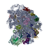
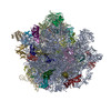
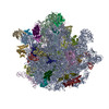
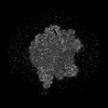
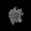
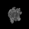
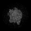
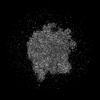
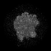
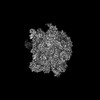
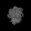
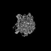
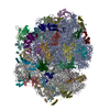
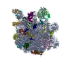
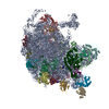
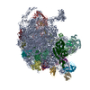
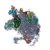
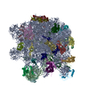
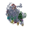
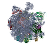
ジャーナル: Proc Natl Acad Sci U S A / 年: 2025 タイトル: HflX-mediated drug resistance through ribosome splitting and rRNA disordering in mycobacteria. 著者: Soneya Majumdar / Amuliya Kashyap / Ravi K Koripella / Manjuli R Sharma / Kelley Hurst-Hess / Swati R Manjari / Nilesh K Banavali / Pallavi Ghosh / Rajendra K Agrawal / 要旨: HflX is a highly conserved ribosome-associated GTPase implicated in rescuing stalled ribosomes and mediating antibiotic resistance in several bacteria, including macrolide-lincosamide antibiotic ...HflX is a highly conserved ribosome-associated GTPase implicated in rescuing stalled ribosomes and mediating antibiotic resistance in several bacteria, including macrolide-lincosamide antibiotic resistance in mycobacteria. Mycobacterial HflXs carry a distinct N-terminal extension (NTE) and a small insertion, as compared to their eubacterial homologs. Here, we present several high-resolution cryo-EM structures of mycobacterial HflX in complex with the 70S ribosome and its 50S subunit, with and without antibiotics. These structures reveal a distinct mechanism for HflX-mediated ribosome splitting and antibiotic resistance in mycobacteria. Our findings indicate that the NTE of mycobacterial HflX induces persistent disordering of multiple 23S rRNA helices, facilitating the dissociation of the 70S ribosome and generating an inactive pool of 50S subunits. During this process, HflX undergoes a large conformational change that stabilizes its NTE. Mycobacterial HflX also acts as an anti-association factor by binding to predissociated 50S subunits. Our structures show that a mycobacteria-specific insertion in HflX reaches far into the peptidyl transferase center (PTC), such that it would overlap with the ribosome-bound macrolide antibiotics. However, in the presence of antibiotics, this insertion retracts, adjusts around, and interacts with the antibiotic molecules. These results suggest that mycobacterial HflX is agnostic to antibiotic presence in the PTC. It mediates antibiotic resistance by splitting antibiotic-stalled 70S ribosomes and inactivating the resulting 50S subunits.
 ムービー
ムービー コントローラー


 データを開く
 基本情報
基本情報
 マップデータ
 試料
試料 キーワード
キーワード 機能・相同性情報
 Mycolicibacterium smegmatis MC2 155 (バクテリア)
 データ登録者
 米国, 3件
  引用
引用 構造の表示
構造の表示
 ダウンロードとリンク
 emd_43333.png
 http://ftp.pdbj.org/pub/emdb/structures/EMD-43333
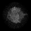


 Z (Sec.)
 Y (Row.)
 X (Col.)


 試料の構成要素
 解析
解析 電子顕微鏡法
 FIELD EMISSION GUN