+ Open data
- Basic information
| Entry |  | |||||||||||||||
|---|---|---|---|---|---|---|---|---|---|---|---|---|---|---|---|---|
| Title | Composite map of AMPylated GlnA bound to hinT | |||||||||||||||
 Map data Map data | composite map of GlnA dimer bound with hinT dimer | |||||||||||||||
 Sample Sample |
| |||||||||||||||
 Keywords Keywords | AMPylation / adenyltransferase / nucleotide binding protein / LIGASE-HYDROLASE complex | |||||||||||||||
| Function / homology |  Function and homology information Function and homology informationammonia assimilation cycle / D-alanine catabolic process / nitrogen utilization / Hydrolases; Acting on phosphorus-nitrogen bonds / adenosine 5'-monophosphoramidase activity / glutamine synthetase / glutamine biosynthetic process / glutamine synthetase activity / response to radiation / nucleotide binding ...ammonia assimilation cycle / D-alanine catabolic process / nitrogen utilization / Hydrolases; Acting on phosphorus-nitrogen bonds / adenosine 5'-monophosphoramidase activity / glutamine synthetase / glutamine biosynthetic process / glutamine synthetase activity / response to radiation / nucleotide binding / protein homodimerization activity / ATP binding / metal ion binding / identical protein binding / membrane / cytoplasm / cytosol Similarity search - Function | |||||||||||||||
| Biological species |  | |||||||||||||||
| Method | single particle reconstruction / cryo EM / Resolution: 2.7 Å | |||||||||||||||
 Authors Authors | Han Y / Sreelatha A / Gonzalez A / Chen Z | |||||||||||||||
| Funding support |  United States, 4 items United States, 4 items
| |||||||||||||||
 Citation Citation |  Journal: To Be Published Journal: To Be PublishedTitle: Composite map of AMPylated GlnA bound to hinT Authors: Gonzalez A / Sreelatha A | |||||||||||||||
| History |
|
- Structure visualization
| Supplemental images |
|---|
- Downloads & links
-EMDB archive
| Map data |  emd_42892.map.gz emd_42892.map.gz | 2.2 MB |  EMDB map data format EMDB map data format | |
|---|---|---|---|---|
| Header (meta data) |  emd-42892-v30.xml emd-42892-v30.xml emd-42892.xml emd-42892.xml | 13.1 KB 13.1 KB | Display Display |  EMDB header EMDB header |
| Images |  emd_42892.png emd_42892.png | 78.2 KB | ||
| Filedesc metadata |  emd-42892.cif.gz emd-42892.cif.gz | 5.8 KB | ||
| Archive directory |  http://ftp.pdbj.org/pub/emdb/structures/EMD-42892 http://ftp.pdbj.org/pub/emdb/structures/EMD-42892 ftp://ftp.pdbj.org/pub/emdb/structures/EMD-42892 ftp://ftp.pdbj.org/pub/emdb/structures/EMD-42892 | HTTPS FTP |
-Validation report
| Summary document |  emd_42892_validation.pdf.gz emd_42892_validation.pdf.gz | 347.8 KB | Display |  EMDB validaton report EMDB validaton report |
|---|---|---|---|---|
| Full document |  emd_42892_full_validation.pdf.gz emd_42892_full_validation.pdf.gz | 347.4 KB | Display | |
| Data in XML |  emd_42892_validation.xml.gz emd_42892_validation.xml.gz | 6 KB | Display | |
| Data in CIF |  emd_42892_validation.cif.gz emd_42892_validation.cif.gz | 6.9 KB | Display | |
| Arichive directory |  https://ftp.pdbj.org/pub/emdb/validation_reports/EMD-42892 https://ftp.pdbj.org/pub/emdb/validation_reports/EMD-42892 ftp://ftp.pdbj.org/pub/emdb/validation_reports/EMD-42892 ftp://ftp.pdbj.org/pub/emdb/validation_reports/EMD-42892 | HTTPS FTP |
-Related structure data
| Related structure data |  8v1yMC  42894  42895 M: atomic model generated by this map C: citing same article ( |
|---|---|
| Similar structure data | Similarity search - Function & homology  F&H Search F&H Search |
- Links
Links
| EMDB pages |  EMDB (EBI/PDBe) / EMDB (EBI/PDBe) /  EMDataResource EMDataResource |
|---|---|
| Related items in Molecule of the Month |
- Map
Map
| File |  Download / File: emd_42892.map.gz / Format: CCP4 / Size: 91.1 MB / Type: IMAGE STORED AS FLOATING POINT NUMBER (4 BYTES) Download / File: emd_42892.map.gz / Format: CCP4 / Size: 91.1 MB / Type: IMAGE STORED AS FLOATING POINT NUMBER (4 BYTES) | ||||||||||||||||||||||||||||||||||||
|---|---|---|---|---|---|---|---|---|---|---|---|---|---|---|---|---|---|---|---|---|---|---|---|---|---|---|---|---|---|---|---|---|---|---|---|---|---|
| Annotation | composite map of GlnA dimer bound with hinT dimer | ||||||||||||||||||||||||||||||||||||
| Projections & slices | Image control
Images are generated by Spider. | ||||||||||||||||||||||||||||||||||||
| Voxel size | X=Y=Z: 0.83 Å | ||||||||||||||||||||||||||||||||||||
| Density |
| ||||||||||||||||||||||||||||||||||||
| Symmetry | Space group: 1 | ||||||||||||||||||||||||||||||||||||
| Details | EMDB XML:
|
-Supplemental data
- Sample components
-Entire : GlnA dimer binding to hinT dimer
| Entire | Name: GlnA dimer binding to hinT dimer |
|---|---|
| Components |
|
-Supramolecule #1: GlnA dimer binding to hinT dimer
| Supramolecule | Name: GlnA dimer binding to hinT dimer / type: complex / ID: 1 / Parent: 0 / Macromolecule list: #1-#2 |
|---|---|
| Source (natural) | Organism:  |
| Molecular weight | Theoretical: 125 KDa |
-Macromolecule #1: Glutamine synthetase
| Macromolecule | Name: Glutamine synthetase / type: protein_or_peptide / ID: 1 / Number of copies: 2 / Enantiomer: LEVO / EC number: glutamine synthetase |
|---|---|
| Source (natural) | Organism:  |
| Molecular weight | Theoretical: 52.57225 KDa |
| Recombinant expression | Organism:  |
| Sequence | String: SEFELMSAEH VLTMLNEHEV KFVDLRFTDT KGKEQHVTIP AHQVNAEFFE EGKMFDGSSI GGWKGINESD MVLMPDASTA VIDPFFADS TLIIRCDILE PGTLQGYDRD PRSIAKRAED YLRSTGIADT VLFGPEPEFF LFDDIRFGSS ISGSHVAIDD I EGAWNSST ...String: SEFELMSAEH VLTMLNEHEV KFVDLRFTDT KGKEQHVTIP AHQVNAEFFE EGKMFDGSSI GGWKGINESD MVLMPDASTA VIDPFFADS TLIIRCDILE PGTLQGYDRD PRSIAKRAED YLRSTGIADT VLFGPEPEFF LFDDIRFGSS ISGSHVAIDD I EGAWNSST QYEGGNKGHR PAVKGGYFPV PPVDSAQDIR SEMCLVMEQM GLVVEAHHHE VATAGQNEVA TRFNTMTKKA DE IQIYKYV VHNVAHRFGK TATFMPKPMF GDNGSGMHCH MSLSKNGVNL FAGDKYAGLS EQALYYIGGV IKHAKAINAL ANP TTNSYK RLVPGYEAPV MLAYSARNRS ASIRIPVVSS PKARRIEVRF PDPAANPYLC FAALLMAGLD GIKNKIHPGE AMDK NLYDL PPEEAKEIPQ VAGSLEEALN ELDLDREFLK AGGVFTDEAI DAYIALRREE DDRVRMTPHP VEFELYYSV UniProtKB: Glutamine synthetase |
-Macromolecule #2: Purine nucleoside phosphoramidase
| Macromolecule | Name: Purine nucleoside phosphoramidase / type: protein_or_peptide / ID: 2 / Number of copies: 2 / Enantiomer: LEVO / EC number: Hydrolases; Acting on phosphorus-nitrogen bonds |
|---|---|
| Source (natural) | Organism:  |
| Molecular weight | Theoretical: 13.637674 KDa |
| Recombinant expression | Organism:  |
| Sequence | String: GAMGSMAEET IFSKIIRREI PSDIVYQDDL VTAFRDISPQ APTHILIIPN ILIPTVNDVS AEHEQALGRM ITVAAKIAEQ EGIAEDGYR LIMNTNRHGG QEVYHINMHL LGGRPLGPML AHKGL UniProtKB: Purine nucleoside phosphoramidase |
-Macromolecule #3: ADENOSINE MONOPHOSPHATE
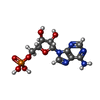
| Macromolecule | Name: ADENOSINE MONOPHOSPHATE / type: ligand / ID: 3 / Number of copies: 2 / Formula: AMP |
|---|---|
| Molecular weight | Theoretical: 347.221 Da |
| Chemical component information |  ChemComp-AMP: |
-Experimental details
-Structure determination
| Method | cryo EM |
|---|---|
 Processing Processing | single particle reconstruction |
| Aggregation state | particle |
- Sample preparation
| Buffer | pH: 7.4 |
|---|---|
| Vitrification | Cryogen name: ETHANE / Instrument: FEI VITROBOT MARK IV |
- Electron microscopy
| Microscope | FEI TITAN KRIOS |
|---|---|
| Image recording | Film or detector model: GATAN K3 BIOQUANTUM (6k x 4k) / Average electron dose: 60.0 e/Å2 |
| Electron beam | Acceleration voltage: 300 kV / Electron source:  FIELD EMISSION GUN FIELD EMISSION GUN |
| Electron optics | Illumination mode: FLOOD BEAM / Imaging mode: BRIGHT FIELD / Nominal defocus max: 2.2 µm / Nominal defocus min: 0.9 µm / Nominal magnification: 105000 |
| Sample stage | Specimen holder model: FEI TITAN KRIOS AUTOGRID HOLDER |
| Experimental equipment |  Model: Titan Krios / Image courtesy: FEI Company |
- Image processing
| Startup model | Type of model: INSILICO MODEL / In silico model: relion ab initio model |
|---|---|
| Final reconstruction | Applied symmetry - Point group: C1 (asymmetric) / Resolution.type: BY AUTHOR / Resolution: 2.7 Å / Resolution method: FSC 0.143 CUT-OFF / Software - Name: RELION (ver. 4.0.1) / Number images used: 225422 |
| Initial angle assignment | Type: MAXIMUM LIKELIHOOD / Software - Name: RELION (ver. 4.0.1) |
| Final angle assignment | Type: MAXIMUM LIKELIHOOD / Software - Name: RELION (ver. 4.0.1) |
-Atomic model buiding 1
| Refinement | Space: REAL |
|---|---|
| Output model |  PDB-8v1y: |
 Movie
Movie Controller


 Z (Sec.)
 Y (Row.)
 X (Col.)


