National Institutes of Health/Office of the Director
DP5OD029613
United States
Swiss National Science Foundation
Switzerland
Human Frontier Science Program (HFSP)
France
Citation
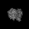
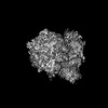
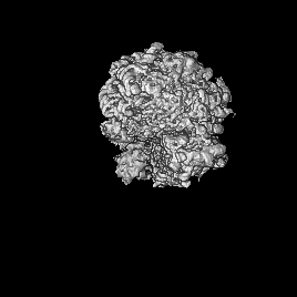
Journal: Nat Struct Mol Biol / Year: 2025 Title: Integrated structural biology of the native malarial translation machinery and its inhibition by an antimalarial drug. Authors: Leonie Anton / Wenjing Cheng / Meseret T Haile / Jerzy M Dziekan / David W Cobb / Xiyan Zhu / Leyan Han / Emerson Li / Anjali Nair / Carolyn L Lee / Hanyu Wang / Hangjun Ke / Guoan Zhang / ...Authors: Leonie Anton / Wenjing Cheng / Meseret T Haile / Jerzy M Dziekan / David W Cobb / Xiyan Zhu / Leyan Han / Emerson Li / Anjali Nair / Carolyn L Lee / Hanyu Wang / Hangjun Ke / Guoan Zhang / Emma H Doud / Alan F Cowman / Chi-Min Ho / Abstract: Our understanding of cellular events is hampered by the gap between the resolution at which we can observe events inside cells and our ability to replicate physiological conditions in test tubes. ...Our understanding of cellular events is hampered by the gap between the resolution at which we can observe events inside cells and our ability to replicate physiological conditions in test tubes. Here, we show in Plasmodium falciparum, a non-model organism of high medical importance, that this gap can be bridged by using an integrated structural biology approach to visualize events inside the cell at molecular resolution. We determined eight high-resolution structures of the native malarial ribosome in actively translating states inside P. falciparum-infected human erythrocytes using in situ cryo-electron tomography. Following perturbation with a Plasmodium-specific translation inhibitor, we then observed a decrease in elongation factor-bound ribosomal states and an apparent upregulation of ribosome biogenesis in inhibitor-treated parasites. Our work elucidates new molecular details of the malarial translation elongation cycle and demonstrates direct multiscale visualization of drug-induced phenotypic changes in the structure and localization of individual molecules within the native cellular context.
In the structure databanks used in Yorodumi, some data are registered as the other names, "COVID-19 virus" and "2019-nCoV". Here are the details of the virus and the list of structure data.
Jan 31, 2019. EMDB accession codes are about to change! (news from PDBe EMDB page)
EMDB accession codes are about to change! (news from PDBe EMDB page)
The allocation of 4 digits for EMDB accession codes will soon come to an end. Whilst these codes will remain in use, new EMDB accession codes will include an additional digit and will expand incrementally as the available range of codes is exhausted. The current 4-digit format prefixed with “EMD-” (i.e. EMD-XXXX) will advance to a 5-digit format (i.e. EMD-XXXXX), and so on. It is currently estimated that the 4-digit codes will be depleted around Spring 2019, at which point the 5-digit format will come into force.
The EM Navigator/Yorodumi systems omit the EMD- prefix.
Related info.:Q: What is EMD? / ID/Accession-code notation in Yorodumi/EM Navigator
Yorodumi is a browser for structure data from EMDB, PDB, SASBDB, etc.
This page is also the successor to EM Navigator detail page, and also detail information page/front-end page for Omokage search.
The word "yorodu" (or yorozu) is an old Japanese word meaning "ten thousand". "mi" (miru) is to see.
Related info.:EMDB / PDB / SASBDB / Comparison of 3 databanks / Yorodumi Search / Aug 31, 2016. New EM Navigator & Yorodumi / Yorodumi Papers / Jmol/JSmol / Function and homology information / Changes in new EM Navigator and Yorodumi
 Movie
Movie Controller

 Yorodumi
 Open data
 Basic information
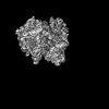
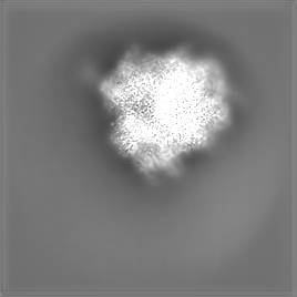
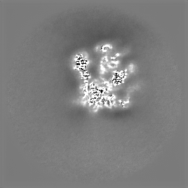
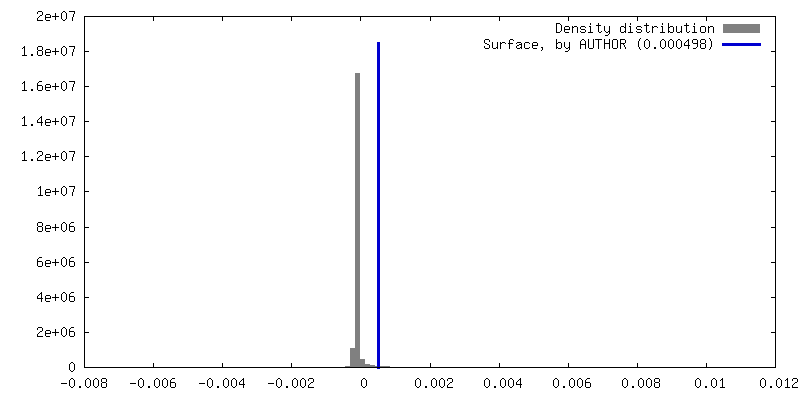
 Map data
 Sample
 Keywords
 Authors
 United States,
  Switzerland,
  France, 3 items
  Citation
 Structure visualization
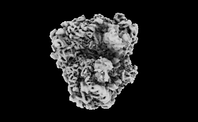
 Downloads & links
 EMDB map data format
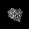
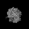
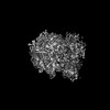
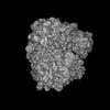
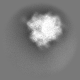
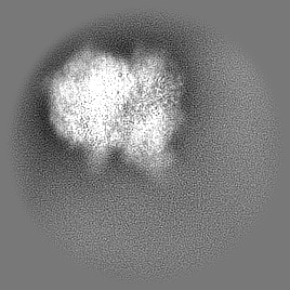
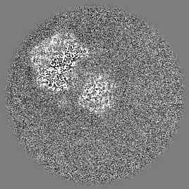
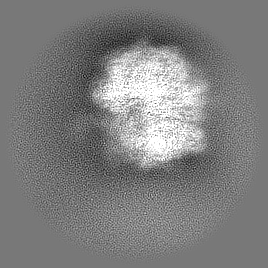
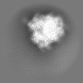
 emd_41494.png
 http://ftp.pdbj.org/pub/emdb/structures/EMD-41494


 Z (Sec.)
 Y (Row.)
 X (Col.)


 Sample components
 Processing
 Electron microscopy
 FIELD EMISSION GUN
