 ムービー
ムービー コントローラー


+ データを開く

- 基本情報
基本情報
| 登録情報 |  | ||||||||||||
|---|---|---|---|---|---|---|---|---|---|---|---|---|---|
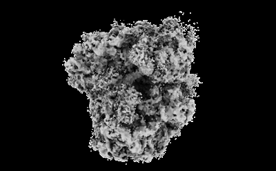
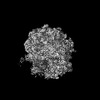
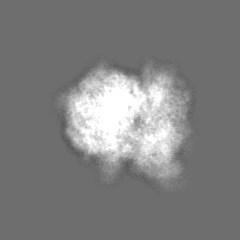
| タイトル | Subtomogram averaged decoding-1 state of the malarial 80S ribosome in Plasmodium falciparum-infected human erythrocytes | ||||||||||||
 マップデータ マップデータ | Decoding-1 translational intermediate from P. falciparum postprocessed | ||||||||||||
 試料 試料 |
| ||||||||||||
 キーワード キーワード | ribosome / malaria / cryoET / in situ / Plasmodium falciparum / TRANSLATION | ||||||||||||
| 生物種 |  | ||||||||||||
| 手法 | サブトモグラム平均法 / クライオ電子顕微鏡法 / 解像度: 6.8 Å | ||||||||||||
 データ登録者 データ登録者 | Anton L / Cheng W / Zhu X / Ho CM | ||||||||||||
| 資金援助 |  米国, 米国,  スイス, スイス,  フランス, 3件 フランス, 3件
| ||||||||||||
 引用 引用 | ジャーナル: To Be Published タイトル: Divergent translational landscape reflects adaptation to biased codon usage in malaria parasites 著者: Anton L / Cheng W / Haile M / Cobb DW / Zhu X / Han L / Li E / Nair A / Lee CL / Ho CM | ||||||||||||
| 履歴 |
|
- 構造の表示
構造の表示
| 添付画像 |
|---|
- ダウンロードとリンク
-EMDBアーカイブ
| マップデータ | emd_41486.map.gz | 7.4 MB |  EMDBマップデータ形式 EMDBマップデータ形式 | |
|---|---|---|---|---|
| ヘッダ (付随情報) | emd-41486-v30.xmlemd-41486.xml | 22.8 KB 22.8 KB | 表示 表示 | EMDBヘッダ |
| FSC (解像度算出) | emd_41486_fsc.xml | 8.6 KB | 表示 | FSCデータファイル |
| 画像 |  emd_41486.png emd_41486.png | 59.6 KB | ||
| Filedesc metadata | emd-41486.cif.gz | 4.1 KB | ||
| その他 | emd_41486_half_map_1.map.gzemd_41486_half_map_2.map.gz | 40.9 MB 40.8 MB | ||
| アーカイブディレクトリ |  http://ftp.pdbj.org/pub/emdb/structures/EMD-41486ftp://ftp.pdbj.org/pub/emdb/structures/EMD-41486 http://ftp.pdbj.org/pub/emdb/structures/EMD-41486ftp://ftp.pdbj.org/pub/emdb/structures/EMD-41486 | HTTPS FTP |
-関連構造データ


-リンク
| EMDBのページ | EMDB (EBI/PDBe) / EMDataResource |
|---|
-マップ
| ファイル | ダウンロード / ファイル: emd_41486.map.gz / 形式: CCP4 / 大きさ: 52.7 MB / タイプ: IMAGE STORED AS FLOATING POINT NUMBER (4 BYTES) | ||||||||||||||||||||||||||||||||||||
|---|---|---|---|---|---|---|---|---|---|---|---|---|---|---|---|---|---|---|---|---|---|---|---|---|---|---|---|---|---|---|---|---|---|---|---|---|---|
| 注釈 | Decoding-1 translational intermediate from P. falciparum postprocessed | ||||||||||||||||||||||||||||||||||||
| 投影像・断面図 | 画像のコントロール
画像は Spider により作成 | ||||||||||||||||||||||||||||||||||||
| ボクセルのサイズ | X=Y=Z: 2.094 Å | ||||||||||||||||||||||||||||||||||||
| 密度 |
| ||||||||||||||||||||||||||||||||||||
| 対称性 | 空間群: 1 | ||||||||||||||||||||||||||||||||||||
| 詳細 | EMDB XML:
|
 Z (Sec.)
 Y (Row.)
 X (Col.)


-添付データ
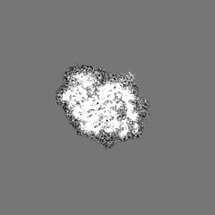
-ハーフマップ: Decoding-1 translational intermediate from P. falciparum half map 1
| ファイル | emd_41486_half_map_1.map | ||||||||||||
|---|---|---|---|---|---|---|---|---|---|---|---|---|---|
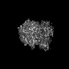
| 注釈 | Decoding-1 translational intermediate from P. falciparum half map 1 | ||||||||||||
| 投影像・断面図 |
| ||||||||||||
| 密度ヒストグラム |


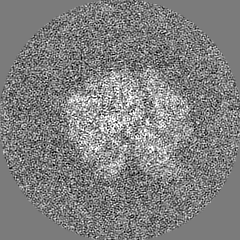
-ハーフマップ: Decoding-1 translational intermediate from P. falciparum half map 2
| ファイル | emd_41486_half_map_2.map | ||||||||||||
|---|---|---|---|---|---|---|---|---|---|---|---|---|---|
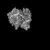
| 注釈 | Decoding-1 translational intermediate from P. falciparum half map 2 | ||||||||||||
| 投影像・断面図 |
| ||||||||||||
| 密度ヒストグラム |


- 試料の構成要素
-全体 : Plasmodium falciparum 80S ribosome consensus structure
| 全体 | 名称: Plasmodium falciparum 80S ribosome consensus structure |
|---|---|
| 要素 |
|
-超分子 #1: Plasmodium falciparum 80S ribosome consensus structure
| 超分子 | 名称: Plasmodium falciparum 80S ribosome consensus structure タイプ: complex / ID: 1 / 親要素: 0 / 含まれる分子: #1-#79 詳細: Subtomogram averaged consensus structure of the malarial 80S ribosome in Plasmodium falciparum-infected human erythrocytes |
|---|---|
| 由来(天然) | 生物種: |
-実験情報
-構造解析
| 手法 | クライオ電子顕微鏡法 |
|---|---|
 解析 解析 | サブトモグラム平均法 |
| 試料の集合状態 | cell |
-試料調製
| 緩衝液 | pH: 7.2 |
|---|---|
| 凍結 | 凍結剤: ETHANE-PROPANE |
- 電子顕微鏡法
| 顕微鏡 | FEI TITAN KRIOS |
|---|---|
| 撮影 | フィルム・検出器のモデル: GATAN K3 (6k x 4k) / 平均電子線量: 2.93 e/Å2 |
| 電子線 | 加速電圧: 300 kV / 電子線源:  FIELD EMISSION GUN FIELD EMISSION GUN |
| 電子光学系 | 照射モード: FLOOD BEAM / 撮影モード: BRIGHT FIELD / 最大 デフォーカス(公称値): 3.2 µm 最小 デフォーカス(公称値): 2.8000000000000003 µm |
| 実験機器 |  モデル: Titan Krios / 画像提供: FEI Company |
-画像解析
| 最終 再構成 | 解像度のタイプ: BY AUTHOR / 解像度: 6.8 Å / 解像度の算出法: FSC 0.143 CUT-OFF / 使用したサブトモグラム数: 7881 |
|---|---|
| 抽出 | トモグラム数: 848 / 使用した粒子像数: 129617 |
| 最終 角度割当 | タイプ: OTHER |
| FSC曲線 (解像度の算出) |  |
