 Movie
Movie Controller


+ Open data

- Basic information
| Entry | Database: EMDB / ID: EMD-4094 | |||||||||
|---|---|---|---|---|---|---|---|---|---|---|
| Title | Structure of a designed Isoaspartyl Dipeptidase filament. | |||||||||
 Map data Map data | ||||||||||
 Sample Sample |
| |||||||||
| Function / homology |  Function and homology information Function and homology informationHydrolases; Acting on peptide bonds (peptidases); Omega peptidases / hydrolase activity, acting on carbon-nitrogen (but not peptide) bonds / beta-aspartyl-peptidase activity / metallopeptidase activity / proteolysis / zinc ion binding / identical protein binding / cytoplasm / cytosol Similarity search - Function | |||||||||
| Biological species |  | |||||||||
| Method | single particle reconstruction / cryo EM / Resolution: 10.5 Å | |||||||||
 Authors Authors | Garcia-Seisdedos H / Empereur-Mot C / Elad N / Levy DE | |||||||||
 Citation Citation | Journal: Nature / Year: 2017 Title: Proteins evolve on the edge of supramolecular self-assembly. Authors: Hector Garcia-Seisdedos / Charly Empereur-Mot / Nadav Elad / Emmanuel D Levy /  Abstract: The self-association of proteins into symmetric complexes is ubiquitous in all kingdoms of life. Symmetric complexes possess unique geometric and functional properties, but their internal symmetry ...The self-association of proteins into symmetric complexes is ubiquitous in all kingdoms of life. Symmetric complexes possess unique geometric and functional properties, but their internal symmetry can pose a risk. In sickle-cell disease, the symmetry of haemoglobin exacerbates the effect of a mutation, triggering assembly into harmful fibrils. Here we examine the universality of this mechanism and its relation to protein structure geometry. We introduced point mutations solely designed to increase surface hydrophobicity among 12 distinct symmetric complexes from Escherichia coli. Notably, all responded by forming supramolecular assemblies in vitro, as well as in vivo upon heterologous expression in Saccharomyces cerevisiae. Remarkably, in four cases, micrometre-long fibrils formed in vivo in response to a single point mutation. Biophysical measurements and electron microscopy revealed that mutants self-assembled in their folded states and so were not amyloid-like. Structural examination of 73 mutants identified supramolecular assembly hot spots predictable by geometry. A subsequent structural analysis of 7,471 symmetric complexes showed that geometric hot spots were buffered chemically by hydrophilic residues, suggesting a mechanism preventing mis-assembly of these regions. Thus, point mutations can frequently trigger folded proteins to self-assemble into higher-order structures. This potential is counterbalanced by negative selection and can be exploited to design nanomaterials in living cells. | |||||||||
| History |
|
- Structure visualization
| Movie |
Movie viewer |
|---|---|
| Structure viewer | EM map: SurfViewMolmilJmol/JSmol |
| Supplemental images |

- Downloads & links
-EMDB archive
| Map data | emd_4094.map.gz | 7.3 MB | EMDB map data format | |
|---|---|---|---|---|
| Header (meta data) | emd-4094-v30.xmlemd-4094.xml | 12.2 KB 12.2 KB | Display Display | EMDB header |
| Images | 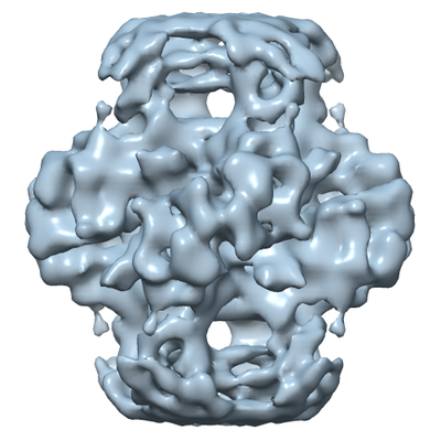 emd_4094.png emd_4094.png | 155.2 KB | ||
| Archive directory |  http://ftp.pdbj.org/pub/emdb/structures/EMD-4094ftp://ftp.pdbj.org/pub/emdb/structures/EMD-4094 http://ftp.pdbj.org/pub/emdb/structures/EMD-4094ftp://ftp.pdbj.org/pub/emdb/structures/EMD-4094 | HTTPS FTP |
-Related structure data
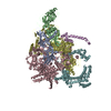
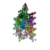
| Related structure data |  5lp3MC M: atomic model generated by this map C: citing same article ( |
|---|---|
| Similar structure data |


-Links
| EMDB pages | EMDB (EBI/PDBe) / EMDataResource |
|---|---|
| Related items in Molecule of the Month |


-Map
| File | Download / File: emd_4094.map.gz / Format: CCP4 / Size: 8 MB / Type: IMAGE STORED AS FLOATING POINT NUMBER (4 BYTES) | ||||||||||||||||||||||||||||||||||||||||||||||||||||||||||||
|---|---|---|---|---|---|---|---|---|---|---|---|---|---|---|---|---|---|---|---|---|---|---|---|---|---|---|---|---|---|---|---|---|---|---|---|---|---|---|---|---|---|---|---|---|---|---|---|---|---|---|---|---|---|---|---|---|---|---|---|---|---|
| Projections & slices | Image control
Images are generated by Spider. | ||||||||||||||||||||||||||||||||||||||||||||||||||||||||||||
| Voxel size | X=Y=Z: 2.11 Å | ||||||||||||||||||||||||||||||||||||||||||||||||||||||||||||
| Density |
| ||||||||||||||||||||||||||||||||||||||||||||||||||||||||||||
| Symmetry | Space group: 1 | ||||||||||||||||||||||||||||||||||||||||||||||||||||||||||||
| Details | EMDB XML:
CCP4 map header:
| ||||||||||||||||||||||||||||||||||||||||||||||||||||||||||||
 Z (Sec.)
 Y (Row.)
 X (Col.)


-Supplemental data
- Sample components
-Entire : Fiber assembly of isoaspartyl dipeptidase E239Y mutant
| Entire | Name: Fiber assembly of isoaspartyl dipeptidase E239Y mutant |
|---|---|
| Components |
|
-Supramolecule #1: Fiber assembly of isoaspartyl dipeptidase E239Y mutant
| Supramolecule | Name: Fiber assembly of isoaspartyl dipeptidase E239Y mutant type: complex / ID: 1 / Parent: 0 / Macromolecule list: all Details: E239Y point mutant induce fiber formation of the isoaspartyl dipeptidase octamers |
|---|---|
| Source (natural) | Organism: |
| Recombinant expression | Organism: Recombinant plasmid: pET-30 a (+) |
-Macromolecule #1: Isoaspartyl Dipeptidase
| Macromolecule | Name: Isoaspartyl Dipeptidase / type: protein_or_peptide / ID: 1 / Enantiomer: LEVO / EC number: beta-aspartyl-peptidase |
|---|---|
| Source (natural) | Organism: |
| Recombinant expression | Organism: |
| Sequence | String: HHHHHHLVPR GIDYTAAGFT LLQGAHLYA PEDRGICDVL V ANGKIIAV ASNIPSDIVP NC TVVDLSG QILCPGFIDQ HVH LIGGGG EAGPTTRTPE VALS RLTEA GVTSVVGLLG TDSIS RHPE SLLAKTRALN EEGISA WML TGAYHVPSRT ITGSVEK DV ...String: HHHHHHLVPR GIDYTAAGFT LLQGAHLYA PEDRGICDVL V ANGKIIAV ASNIPSDIVP NC TVVDLSG QILCPGFIDQ HVH LIGGGG EAGPTTRTPE VALS RLTEA GVTSVVGLLG TDSIS RHPE SLLAKTRALN EEGISA WML TGAYHVPSRT ITGSVEK DV AIIDRVIGVK CAISDHRS A APDVYHLANM AAESRVGGL LGGKPGVTVF HMGDSKKALQ PIYDLLENC DVPISKLLPT H VNRNVPLF YQALEFARKG GT IDITSSI DEPVAPAEGI ARA VQAGIP LARVTLSSDG NGSQ PFFDD EGNLTHIGVA GFETL LETV QVLVKDYDFS ISDALR PLT SSVAGFLNLT GKGEILP GN DADLLVMTPE LRIEQVYA R GKLMVKDGKA CVKGTFETA |
-Experimental details
-Structure determination
| Method | cryo EM |
|---|---|
 Processing Processing | single particle reconstruction |
| Aggregation state | filament |
-Sample preparation
| Concentration | 0.2 mg/mL | |||||||||
|---|---|---|---|---|---|---|---|---|---|---|
| Buffer | pH: 7.5 Component:
| |||||||||
| Grid | Model: Quantifoil / Material: COPPER / Mesh: 300 / Support film - Material: CARBON / Support film - topology: CONTINUOUS / Pretreatment - Type: GLOW DISCHARGE / Pretreatment - Atmosphere: AIR | |||||||||
| Vitrification | Cryogen name: ETHANE / Chamber humidity: 95 % / Chamber temperature: 297 K / Instrument: LEICA EM GP |
- Electron microscopy
| Microscope | FEI TECNAI F20 |
|---|---|
| Image recording | Film or detector model: GATAN K2 SUMMIT (4k x 4k) / Average electron dose: 32.0 e/Å2 |
| Electron beam | Acceleration voltage: 200 kV / Electron source:  FIELD EMISSION GUN FIELD EMISSION GUN |
| Electron optics | C2 aperture diameter: 30.0 µm / Illumination mode: FLOOD BEAM / Imaging mode: BRIGHT FIELD |
| Experimental equipment |  Model: Tecnai F20 / Image courtesy: FEI Company |
+Image processing

-Atomic model buiding 1
| Refinement | Protocol: RIGID BODY FIT |
|---|---|
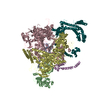
| Output model | PDB-5lp3: |
