 Movie
Movie Controller

[English] 日本語
 Yorodumi
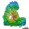
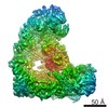
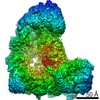
- EMDB-4047: Cryo-EM structure of ternary deletion mutant of human APC/C-Cdh1-... -
+ Open data

- Basic information
| Entry | Database: EMDB / ID: EMD-4047 | |||||||||
|---|---|---|---|---|---|---|---|---|---|---|
| Title | Cryo-EM structure of ternary deletion mutant of human APC/C-Cdh1-Hsl1 complex without Apc1 WD40 domain | |||||||||
 Map data Map data | ||||||||||
 Sample Sample |
| |||||||||
| Biological species |  Homo sapiens (human) Homo sapiens (human) | |||||||||
| Method | single particle reconstruction / cryo EM / Resolution: 6.0 Å | |||||||||
 Authors Authors | Li Q / Chang L / Yang J / Zhang Z / Barford D | |||||||||
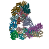
 Citation Citation | Journal: Proc Natl Acad Sci U S A / Year: 2016 Title: WD40 domain of Apc1 is critical for the coactivator-induced allosteric transition that stimulates APC/C catalytic activity. Authors: Qiuhong Li / Leifu Chang / Shintaro Aibara / Jing Yang / Ziguo Zhang / David Barford /  Abstract: The anaphase-promoting complex/cyclosome (APC/C) is a large multimeric cullin-RING E3 ubiquitin ligase that orchestrates cell-cycle progression by targeting cell-cycle regulatory proteins for ...The anaphase-promoting complex/cyclosome (APC/C) is a large multimeric cullin-RING E3 ubiquitin ligase that orchestrates cell-cycle progression by targeting cell-cycle regulatory proteins for destruction via the ubiquitin proteasome system. The APC/C assembly comprises two scaffolding subcomplexes: the platform and the TPR lobe that together coordinate the juxtaposition of the catalytic and substrate-recognition modules. The platform comprises APC/C subunits Apc1, Apc4, Apc5, and Apc15. Although the role of Apc1 as an APC/C scaffolding subunit has been characterized, its specific functions in contributing toward APC/C catalytic activity are not fully understood. Here, we report the crystal structure of the N-terminal domain of human Apc1 (Apc1N) determined at 2.2-Å resolution and provide an atomic-resolution description of the architecture of its WD40 (WD40 repeat) domain (Apc1(WD40)). To understand how Apc1(WD40) contributes to APC/C activity, a mutant form of the APC/C with Apc1(WD40) deleted was generated and evaluated biochemically and structurally. We found that the deletion of Apc1(WD40) abolished the UbcH10-dependent ubiquitination of APC/C substrates without impairing the Ube2S-dependent ubiquitin chain elongation activity. A cryo-EM structure of an APC/C-Cdh1 complex with Apc1(WD40) deleted showed that the mutant APC/C is locked into an inactive conformation in which the UbcH10-binding site of the catalytic module is inaccessible. Additionally, an EM density for Apc15 is not visible. Our data show that Apc1(WD40) is required to mediate the coactivator-induced conformational change of the APC/C that is responsible for stimulating APC/C catalytic activity by promoting UbcH10 binding. In contrast, Ube2S activity toward APC/C substrates is not dependent on the initiation-competent conformation of the APC/C. | |||||||||
| History |
|
- Structure visualization
| Movie |
 Movie viewer Movie viewer |
|---|---|
| Structure viewer | EM map: SurfViewMolmilJmol/JSmol |
| Supplemental images |

- Downloads & links
-EMDB archive
| Map data | emd_4047.map.gz | 7 MB | EMDB map data format | |
|---|---|---|---|---|
| Header (meta data) | emd-4047-v30.xmlemd-4047.xml | 12.4 KB 12.4 KB | Display Display | EMDB header |
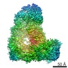
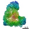
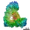
| Images | 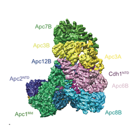 emd_4047.png emd_4047.png | 75 KB | ||
| Archive directory |  http://ftp.pdbj.org/pub/emdb/structures/EMD-4047ftp://ftp.pdbj.org/pub/emdb/structures/EMD-4047 http://ftp.pdbj.org/pub/emdb/structures/EMD-4047ftp://ftp.pdbj.org/pub/emdb/structures/EMD-4047 | HTTPS FTP |
-Related structure data


-Links
| EMDB pages | EMDB (EBI/PDBe) / EMDataResource |
|---|
-Map
| File | Download / File: emd_4047.map.gz / Format: CCP4 / Size: 70.2 MB / Type: IMAGE STORED AS FLOATING POINT NUMBER (4 BYTES) | ||||||||||||||||||||||||||||||||||||||||||||||||||||||||||||
|---|---|---|---|---|---|---|---|---|---|---|---|---|---|---|---|---|---|---|---|---|---|---|---|---|---|---|---|---|---|---|---|---|---|---|---|---|---|---|---|---|---|---|---|---|---|---|---|---|---|---|---|---|---|---|---|---|---|---|---|---|---|
| Projections & slices | Image control
Images are generated by Spider. | ||||||||||||||||||||||||||||||||||||||||||||||||||||||||||||
| Voxel size | X=Y=Z: 1.36 Å | ||||||||||||||||||||||||||||||||||||||||||||||||||||||||||||
| Density |
| ||||||||||||||||||||||||||||||||||||||||||||||||||||||||||||
| Symmetry | Space group: 1 | ||||||||||||||||||||||||||||||||||||||||||||||||||||||||||||
| Details | EMDB XML:
CCP4 map header:
| ||||||||||||||||||||||||||||||||||||||||||||||||||||||||||||
 Z (Sec.)
 Y (Row.)
 X (Col.)


-Supplemental data
- Sample components
-Entire : Ternary complex of APC/C-Cdh1-Hsl1 without Apc1 WD40 domain
| Entire | Name: Ternary complex of APC/C-Cdh1-Hsl1 without Apc1 WD40 domain |
|---|---|
| Components |
|
-Supramolecule #1: Ternary complex of APC/C-Cdh1-Hsl1 without Apc1 WD40 domain
| Supramolecule | Name: Ternary complex of APC/C-Cdh1-Hsl1 without Apc1 WD40 domain type: complex / ID: 1 / Parent: 0 |
|---|---|
| Source (natural) | Organism: Homo sapiens (human) |
| Recombinant expression | Organism:  Trichoplusia ni (cabbage looper) / Recombinant plasmid: pFU Trichoplusia ni (cabbage looper) / Recombinant plasmid: pFU |
| Molecular weight | Theoretical: 1.2 MDa |
-Experimental details
-Structure determination
| Method | cryo EM |
|---|---|
 Processing Processing | single particle reconstruction |
| Aggregation state | cell |
-Sample preparation
| Concentration | 0.12 mg/mL | ||||||||
|---|---|---|---|---|---|---|---|---|---|
| Buffer | pH: 8 Component:
Details: Solutions were made fresh from powder and filtered to avoid microbial contamination. | ||||||||
| Grid | Model: Quantifoil R2/2 / Material: COPPER / Mesh: 300 / Support film - Material: CARBON / Support film - topology: HOLEY ARRAY / Support film - Film thickness: 30.0 nm / Pretreatment - Type: PLASMA CLEANING / Pretreatment - Atmosphere: OTHER Details: The grid was coated with continuous carbon film prior to use. | ||||||||
| Vitrification | Cryogen name: ETHANE / Chamber humidity: 100 % / Chamber temperature: 277 K / Instrument: FEI VITROBOT MARK III / Details: Blot for 5 seconds before plunging.. | ||||||||
| Details | Recombinant protein complex expressed and purified from insect cells. This sample was monodisperse. |
- Electron microscopy
| Microscope | FEI POLARA 300 |
|---|---|
| Image recording | Film or detector model: FEI FALCON II (4k x 4k) / Detector mode: OTHER / Digitization - Dimensions - Width: 4096 pixel / Digitization - Dimensions - Height: 4096 pixel / Number grids imaged: 4 / Number real images: 1729 / Average exposure time: 1.1 sec. / Average electron dose: 14.6 e/Å2 |
| Electron beam | Acceleration voltage: 300 kV / Electron source:  FIELD EMISSION GUN FIELD EMISSION GUN |
| Electron optics | Calibrated defocus max: 4.0 µm / Calibrated defocus min: 1.5 µm / Illumination mode: OTHER / Imaging mode: BRIGHT FIELD / Cs: 2.0 mm |
| Experimental equipment |  Model: Tecnai Polara / Image courtesy: FEI Company |
+Image processing
-Atomic model buiding 1
| Refinement | Space: REAL / Protocol: FLEXIBLE FIT |
|---|
