Movie
Movie Controller

[English] 日本語
 Yorodumi
- EMDB-39732: Cryo-EM structure of apo Aspergillus niger glutamate dehydrogenas... -
+ Open data

- Basic information
| Entry |  | |||||||||
|---|---|---|---|---|---|---|---|---|---|---|
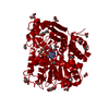
| Title | Cryo-EM structure of apo Aspergillus niger glutamate dehydrogenase (AnGDH) in the closed conformation | |||||||||
 Map data Map data | AnGDH closed conformation cryo-EM map | |||||||||
 Sample Sample |
| |||||||||
 Keywords Keywords | glutamate dehydrogenase / allostery / cooperativity / Aspergillus / cryo-EM / domain dynamics / OXIDOREDUCTASE | |||||||||
| Function / homology |  Function and homology information Function and homology informationL-glutamate dehydrogenase (NADP+) activity / : / nucleotide binding / cytosol Similarity search - Function | |||||||||
| Biological species |  | |||||||||
| Method | single particle reconstruction / cryo EM / Resolution: 3.6 Å | |||||||||
 Authors Authors | Godsora BKJ / Das P / Bhaumik P | |||||||||
| Funding support |  India, 1 items India, 1 items
| |||||||||
 Citation Citation | Journal: Protein Sci / Year: 2025 Title: Conformational flexibility associated with remote residues regulates the kinetic properties of glutamate dehydrogenase. Authors: Barsa Kanchan Jyotshna Godsora / Parijat Das / Prasoon Kumar Mishra / Anjali Sairaman / Sandip Kaledhonkar / Narayan S Punekar / Prasenjit Bhaumik / Abstract: Glutamate dehydrogenase (GDH) is a pivotal metabolic enzyme in all living organisms, and some of the GDHs exhibit substrate-dependent homotropic cooperativity. However, the mode of allosteric ...Glutamate dehydrogenase (GDH) is a pivotal metabolic enzyme in all living organisms, and some of the GDHs exhibit substrate-dependent homotropic cooperativity. However, the mode of allosteric communication during the homotropic effect in GDHs remains poorly understood. In this study, we examined two homologous GDHs, Aspergillus niger GDH (AnGDH) and Aspergillus terreus GDH (AtGDH), with differing substrate utilization kinetics to uncover the factors driving their distinct behavior. We report the crystal structures and first-ever cryo-EM structures of apo- AtGDH and AnGDH that captured arrays of conformational ensembles. A wider mouth opening (~ 21 Å) is observed for the cooperative AnGDH as compared to the non-cooperative AtGDH (~17 Å) in their apo states. A network of interactions related to the substitutions in Domain II influence structural flexibility in these GDHs. Remarkably, we have identified a distant substitution (R246 to S) in Domain II, as a part of this network, which can impact the mouth opening and converts non-cooperative AtGDH into a cooperative enzyme. Our study demonstrates that remote residues can influence structural and kinetic properties in homologous GDHs. | |||||||||
| History |
|
- Structure visualization
| Supplemental images |
|---|

- Downloads & links
-EMDB archive
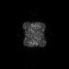
| Map data | emd_39732.map.gz | 49.3 MB | EMDB map data format | |
|---|---|---|---|---|
| Header (meta data) | emd-39732-v30.xmlemd-39732.xml | 16.1 KB 16.1 KB | Display Display | EMDB header |
| Images |  emd_39732.png emd_39732.png | 91.7 KB | ||
| Filedesc metadata | emd-39732.cif.gz | 5.7 KB | ||
| Others | emd_39732_half_map_1.map.gzemd_39732_half_map_2.map.gz | 40.5 MB 40.5 MB | ||
| Archive directory |  http://ftp.pdbj.org/pub/emdb/structures/EMD-39732ftp://ftp.pdbj.org/pub/emdb/structures/EMD-39732 http://ftp.pdbj.org/pub/emdb/structures/EMD-39732ftp://ftp.pdbj.org/pub/emdb/structures/EMD-39732 | HTTPS FTP |
-Related structure data
| Related structure data |  8z1oMC  8z1cC  8z1mC  8z1nC  8z29C  8z2aC  8z2bC  8z2cC  8z2fC M: atomic model generated by this map C: citing same article ( |
|---|---|
| Similar structure data |


-Links
| EMDB pages | EMDB (EBI/PDBe) / EMDataResource |
|---|
-Map
| File | Download / File: emd_39732.map.gz / Format: CCP4 / Size: 52.7 MB / Type: IMAGE STORED AS FLOATING POINT NUMBER (4 BYTES) | ||||||||||||||||||||||||||||||||||||
|---|---|---|---|---|---|---|---|---|---|---|---|---|---|---|---|---|---|---|---|---|---|---|---|---|---|---|---|---|---|---|---|---|---|---|---|---|---|
| Annotation | AnGDH closed conformation cryo-EM map | ||||||||||||||||||||||||||||||||||||
| Projections & slices | Image control
Images are generated by Spider. | ||||||||||||||||||||||||||||||||||||
| Voxel size | X=Y=Z: 1.38 Å | ||||||||||||||||||||||||||||||||||||
| Density |
| ||||||||||||||||||||||||||||||||||||
| Symmetry | Space group: 1 | ||||||||||||||||||||||||||||||||||||
| Details | EMDB XML:
|
 Z (Sec.)
 Y (Row.)
 X (Col.)


-Supplemental data
-Half map: AnGDH closed conformation half1 map
| File | emd_39732_half_map_1.map | ||||||||||||
|---|---|---|---|---|---|---|---|---|---|---|---|---|---|
| Annotation | AnGDH closed conformation half1 map | ||||||||||||
| Projections & Slices |
| ||||||||||||
| Density Histograms |


-Half map: AnGDH closed conformation half2 map
| File | emd_39732_half_map_2.map | ||||||||||||
|---|---|---|---|---|---|---|---|---|---|---|---|---|---|
| Annotation | AnGDH closed conformation half2 map | ||||||||||||
| Projections & Slices |
| ||||||||||||
| Density Histograms |


- Sample components
-Entire : apo form of Aspergillus niger glutamate dehydrogenase
| Entire | Name: apo form of Aspergillus niger glutamate dehydrogenase |
|---|---|
| Components |
|
-Supramolecule #1: apo form of Aspergillus niger glutamate dehydrogenase
| Supramolecule | Name: apo form of Aspergillus niger glutamate dehydrogenase / type: complex / ID: 1 / Parent: 0 / Macromolecule list: all |
|---|---|
| Source (natural) | Organism: |
| Molecular weight | Theoretical: 300 KDa |
-Macromolecule #1: Glutamate dehydrogenase
| Macromolecule | Name: Glutamate dehydrogenase / type: protein_or_peptide / ID: 1 / Number of copies: 6 / Enantiomer: LEVO |
|---|---|
| Source (natural) | Organism: |
| Molecular weight | Theoretical: 49.442508 KDa |
| Recombinant expression | Organism:  |
| Sequence | String: MSNLPHEPEF EQAYKELAST LENSTLFQKN PEYRKALAVV SVPERVIQFR VVWEDDAGNV QVNRGFRVQF NSALGPYKGG LRFHPSVNL SILKFLGFEQ IFKNALTGLN MGGGKGGSDF DPKGKSDNEI RRFCVSFMTE LCKHIGADTD VPAGDIGVTG R EVGFLFGQ ...String: MSNLPHEPEF EQAYKELAST LENSTLFQKN PEYRKALAVV SVPERVIQFR VVWEDDAGNV QVNRGFRVQF NSALGPYKGG LRFHPSVNL SILKFLGFEQ IFKNALTGLN MGGGKGGSDF DPKGKSDNEI RRFCVSFMTE LCKHIGADTD VPAGDIGVTG R EVGFLFGQ YRKIRNQWEG VLTGKGGSWG GSLIRPEATG YGVVYYVEHM IAHATNGQES FKGKRVAISG SGNVAQYAAL KV IELGGSV VSLSDSQGSL IINGEGSFTP EEIELIAQTK VERKQLASIV GAAPFSDANK FKYIAGARPW VHVGKVDVAL PSA TQNEIS GEEAQVLINA GCKFIAEGSN MGCTQEAIDT FEAHRTANAG AAAIWYAPGK AANAGGVAVS GLEMAQNSAR LSWT SEEVD ARLKDIMRDC FKNGLETAQE YATPAEGVLP SLVTGSNIAG FTKVAAAMKD QGDWW UniProtKB: Glutamate dehydrogenase |
-Experimental details
-Structure determination
| Method | cryo EM |
|---|---|
 Processing Processing | single particle reconstruction |
| Aggregation state | particle |
-Sample preparation
| Concentration | 6 mg/mL |
|---|---|
| Buffer | pH: 7.5 / Details: 30 mM Phosphate buffer pH 7.5 |
| Grid | Model: Quantifoil R0.6/1 / Material: GOLD / Mesh: 300 / Pretreatment - Type: GLOW DISCHARGE |
| Vitrification | Cryogen name: ETHANE |
- Electron microscopy
| Microscope | FEI TITAN KRIOS |
|---|---|
| Image recording | Film or detector model: FEI FALCON III (4k x 4k) / Detector mode: COUNTING / Average electron dose: 27.67 e/Å2 |
| Electron beam | Acceleration voltage: 300 kV / Electron source:  FIELD EMISSION GUN FIELD EMISSION GUN |
| Electron optics | Illumination mode: SPOT SCAN / Imaging mode: BRIGHT FIELD / Nominal defocus max: 3.3000000000000003 µm / Nominal defocus min: 2.1 µm |
| Experimental equipment |  Model: Titan Krios / Image courtesy: FEI Company |
-Image processing
| Startup model | Type of model: PDB ENTRY PDB model - PDB ID: |
|---|---|
| Final reconstruction | Number classes used: 1 / Applied symmetry - Point group: D3 (2x3 fold dihedral) / Resolution.type: BY AUTHOR / Resolution: 3.6 Å / Resolution method: FSC 0.143 CUT-OFF / Number images used: 207704 |
| Initial angle assignment | Type: NOT APPLICABLE |
| Final angle assignment | Type: NOT APPLICABLE |