 ムービー
ムービー コントローラー


+ データを開く

- 基本情報
基本情報
| 登録情報 | データベース: EMDB / ID: EMD-3874 | ||||||||||||
|---|---|---|---|---|---|---|---|---|---|---|---|---|---|
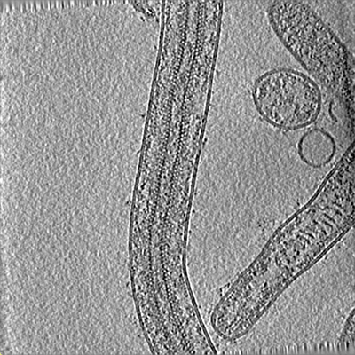
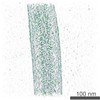
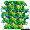
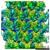
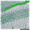
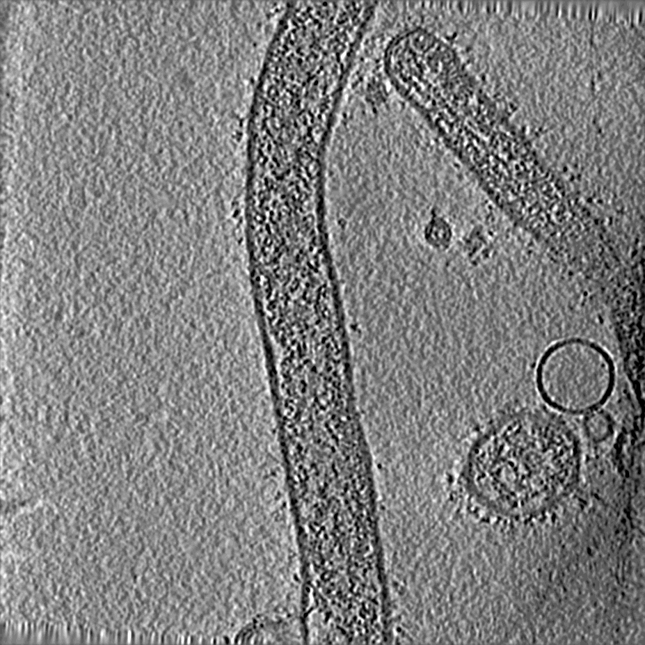
| タイトル | Cryo-electron tomogram of Ebola virus | ||||||||||||
 マップデータ マップデータ | A 4x binned representative tomogram of intact Ebola virus virions | ||||||||||||
 試料 試料 |
| ||||||||||||
| 生物種 |   Ebola virus - Mayinga, Zaire, 1976 (エボラウイルス) Ebola virus - Mayinga, Zaire, 1976 (エボラウイルス) | ||||||||||||
| 手法 | 電子線トモグラフィー法 / クライオ電子顕微鏡法 | ||||||||||||
 データ登録者 データ登録者 | Wan W / Kolesnikova L / Clarke M / Koehler A / Noda T / Becker S / Briggs JAG | ||||||||||||
| 資金援助 |  ドイツ, 3件 ドイツ, 3件
| ||||||||||||
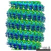
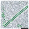
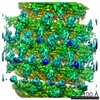
 引用 引用 | ジャーナル: Nature / 年: 2017 タイトル: Structure and assembly of the Ebola virus nucleocapsid. 著者: William Wan / Larissa Kolesnikova / Mairi Clarke / Alexander Koehler / Takeshi Noda / Stephan Becker / John A G Briggs /   要旨: Ebola and Marburg viruses are filoviruses: filamentous, enveloped viruses that cause haemorrhagic fever. Filoviruses are within the order Mononegavirales, which also includes rabies virus, measles ...Ebola and Marburg viruses are filoviruses: filamentous, enveloped viruses that cause haemorrhagic fever. Filoviruses are within the order Mononegavirales, which also includes rabies virus, measles virus, and respiratory syncytial virus. Mononegaviruses have non-segmented, single-stranded negative-sense RNA genomes that are encapsidated by nucleoprotein and other viral proteins to form a helical nucleocapsid. The nucleocapsid acts as a scaffold for virus assembly and as a template for genome transcription and replication. Insights into nucleoprotein-nucleoprotein interactions have been derived from structural studies of oligomerized, RNA-encapsidating nucleoprotein, and cryo-electron microscopy of nucleocapsid or nucleocapsid-like structures. There have been no high-resolution reconstructions of complete mononegavirus nucleocapsids. Here we apply cryo-electron tomography and subtomogram averaging to determine the structure of Ebola virus nucleocapsid within intact viruses and recombinant nucleocapsid-like assemblies. These structures reveal the identity and arrangement of the nucleocapsid components, and suggest that the formation of an extended α-helix from the disordered carboxy-terminal region of nucleoprotein-core links nucleoprotein oligomerization, nucleocapsid condensation, RNA encapsidation, and accessory protein recruitment. | ||||||||||||
| 履歴 |
|
- 構造の表示
構造の表示
| ムービー |
 ムービービューア ムービービューア |
|---|---|
| 構造ビューア | EMマップ: SurfViewMolmilJmol/JSmol |
| 添付画像 |
- ダウンロードとリンク
-EMDBアーカイブ
| マップデータ | emd_3874.map.gz | 80 MB | EMDBマップデータ形式 | |
|---|---|---|---|---|
| ヘッダ (付随情報) | emd-3874-v30.xmlemd-3874.xml | 12.9 KB 12.9 KB | 表示 表示 | EMDBヘッダ |
| 画像 |  emd_3874.png emd_3874.png | 224.4 KB | ||
| アーカイブディレクトリ |  http://ftp.pdbj.org/pub/emdb/structures/EMD-3874ftp://ftp.pdbj.org/pub/emdb/structures/EMD-3874 http://ftp.pdbj.org/pub/emdb/structures/EMD-3874ftp://ftp.pdbj.org/pub/emdb/structures/EMD-3874 | HTTPS FTP |
-検証レポート
| 文書・要旨 | emd_3874_validation.pdf.gz | 200.2 KB | 表示 | EMDB検証レポート |
|---|---|---|---|---|
| 文書・詳細版 | emd_3874_full_validation.pdf.gz | 199.3 KB | 表示 | |
| XML形式データ | emd_3874_validation.xml.gz | 3.2 KB | 表示 | |
| アーカイブディレクトリ | https://ftp.pdbj.org/pub/emdb/validation_reports/EMD-3874ftp://ftp.pdbj.org/pub/emdb/validation_reports/EMD-3874 | HTTPS FTP |
-関連構造データ
-リンク
| EMDBのページ | EMDB (EBI/PDBe) / EMDataResource |
|---|
-マップ
| ファイル | ダウンロード / ファイル: emd_3874.map.gz / 形式: CCP4 / 大きさ: 409.8 MB / タイプ: IMAGE STORED AS SIGNED INTEGER (2 BYTES) | ||||||||||||||||||||||||||||||||||||||||||||||||||||||||||||||||||||
|---|---|---|---|---|---|---|---|---|---|---|---|---|---|---|---|---|---|---|---|---|---|---|---|---|---|---|---|---|---|---|---|---|---|---|---|---|---|---|---|---|---|---|---|---|---|---|---|---|---|---|---|---|---|---|---|---|---|---|---|---|---|---|---|---|---|---|---|---|---|
| 注釈 | A 4x binned representative tomogram of intact Ebola virus virions | ||||||||||||||||||||||||||||||||||||||||||||||||||||||||||||||||||||
| 投影像・断面図 | 画像のコントロール
画像は Spider により作成 これらの図は立方格子座標系で作成されたものです | ||||||||||||||||||||||||||||||||||||||||||||||||||||||||||||||||||||
| ボクセルのサイズ | X=Y=Z: 7.12 Å | ||||||||||||||||||||||||||||||||||||||||||||||||||||||||||||||||||||
| 密度 |
| ||||||||||||||||||||||||||||||||||||||||||||||||||||||||||||||||||||
| 対称性 | 空間群: 1 | ||||||||||||||||||||||||||||||||||||||||||||||||||||||||||||||||||||
| 詳細 | EMDB XML:
CCP4マップ ヘッダ情報:
| ||||||||||||||||||||||||||||||||||||||||||||||||||||||||||||||||||||
 Z (Sec.)
 Y (Row.)
 X (Col.)


-添付データ
- 試料の構成要素
-全体 : Ebola virus - Mayinga, Zaire, 1976
| 全体 | 名称: Ebola virus - Mayinga, Zaire, 1976 (エボラウイルス) |
|---|---|
| 要素 |
|
-超分子 #1: Ebola virus - Mayinga, Zaire, 1976
| 超分子 | 名称: Ebola virus - Mayinga, Zaire, 1976 / タイプ: virus / ID: 1 / 親要素: 0 / 含まれる分子: #1-#3 詳細: Virus was isolated from infected VeroE6 cells. Purified viruses were fixed with paraformaldehyde. NCBI-ID: 128952 / 生物種: Ebola virus - Mayinga, Zaire, 1976 / ウイルスタイプ: VIRION / ウイルス・単離状態: STRAIN / ウイルス・エンベロープ: Yes / ウイルス・中空状態: No |
|---|---|
| ウイルス殻 | Shell ID: 1 / 名称: Nucleocapsid / 直径: 280.0 Å |
-実験情報
-構造解析
| 手法 | クライオ電子顕微鏡法 |
|---|---|
 解析 解析 | 電子線トモグラフィー法 |
| 試料の集合状態 | helical array |
-試料調製
| 緩衝液 | pH: 7.4 詳細: Virus was purified into Dulbecco's modified Eagle's medium (DMEM) with 4% paraformaldehyde |
|---|---|
| グリッド | モデル: C-flat 2/1 3C / 材質: COPPER / メッシュ: 300 / 支持フィルム - 材質: CARBON / 支持フィルム - トポロジー: HOLEY / 支持フィルム - Film thickness: 20.0 nm / 前処理 - タイプ: GLOW DISCHARGE / 前処理 - 雰囲気: AIR / 前処理 - 気圧: 0.039 kPa |
| 凍結 | 凍結剤: ETHANE / チャンバー内湿度: 95 % / 装置: FEI VITROBOT MARK II |
| 切片作成 | その他: NO SECTIONING |
| 位置合わせマーカー | Manufacturer: UMC Utrecht / 直径: 10 nm |
- 電子顕微鏡法
| 顕微鏡 | FEI TITAN KRIOS |
|---|---|
| 特殊光学系 | エネルギーフィルター - 名称: GIF Quantum LS エネルギーフィルター - エネルギー下限: -10 eV エネルギーフィルター - エネルギー上限: 10 eV |
| 撮影 | フィルム・検出器のモデル: GATAN K2 QUANTUM (4k x 4k) 検出モード: SUPER-RESOLUTION / デジタル化 - サイズ - 横: 3708 pixel / デジタル化 - サイズ - 縦: 3708 pixel / デジタル化 - 画像ごとのフレーム数: 1-5 / 平均露光時間: 2.0 sec. / 平均電子線量: 2.9 e/Å2 |
| 電子線 | 加速電圧: 300 kV / 電子線源:  FIELD EMISSION GUN FIELD EMISSION GUN |
| 電子光学系 | C2レンズ絞り径: 50.0 µm / 照射モード: FLOOD BEAM / 撮影モード: BRIGHT FIELD / Cs: 2.7 mm / 最大 デフォーカス(公称値): 4.5 µm / 最小 デフォーカス(公称値): 2.0 µm / 倍率(公称値): 81000 |
| 試料ステージ | 試料ホルダーモデル: FEI TITAN KRIOS AUTOGRID HOLDER ホルダー冷却材: NITROGEN |
| 実験機器 |  モデル: Titan Krios / 画像提供: FEI Company |
-画像解析
| 詳細 | Frames were aligned using K2Align software, based off the MotionCorr algorithm. Tomograms were reconstructed with IMOD, using stripwise CTF-correction and weighted back projection. | ||||||
|---|---|---|---|---|---|---|---|
| 最終 再構成 | アルゴリズム: BACK PROJECTION / ソフトウェア - 名称: IMOD / 使用した粒子像数: 41 | ||||||
| CTF補正 | ソフトウェア:
詳細: CTF amplitude correction was performed during the wedge-weighted subtomogram averaging step. |
