Movie
Movie Controller

[English] 日本語
 Yorodumi
- EMDB-3843: Structure of a native assembly intermediate of the human mitochon... -
+ Open data

- Basic information
| Entry | Database: EMDB / ID: EMD-3843 | |||||||||
|---|---|---|---|---|---|---|---|---|---|---|
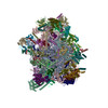
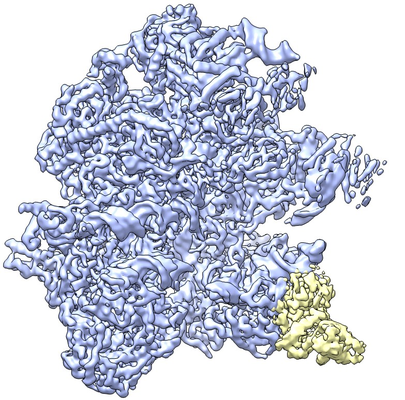
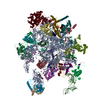
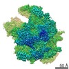
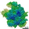
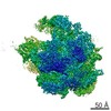
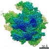
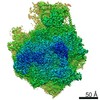
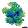
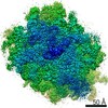
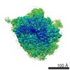
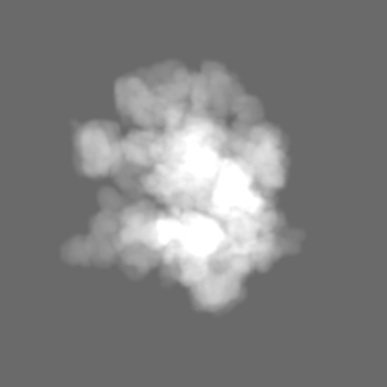
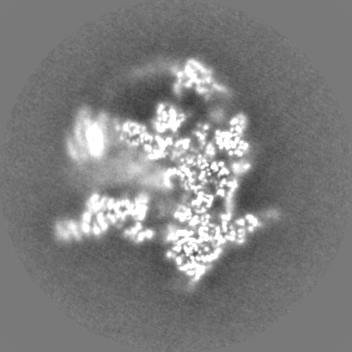
| Title | Structure of a native assembly intermediate of the human mitochondrial ribosome with unfolded interfacial rRNA | |||||||||
 Map data Map data | ||||||||||
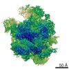
 Sample Sample |
| |||||||||
 Keywords Keywords | Ribosome / mitochondria / biogenesis / translation / electron cryomicroscopy | |||||||||
| Function / homology |  Function and homology information Function and homology informationnegative regulation of mitochondrial translation / negative regulation of ribosome biogenesis / Mitochondrial Fatty Acid Beta-Oxidation / Protein lipoylation / positive regulation of mitochondrial translation / Complex I biogenesis / Respiratory electron transport / mitochondrial large ribosomal subunit assembly / rRNA import into mitochondrion / mitochondrial translational termination ...negative regulation of mitochondrial translation / negative regulation of ribosome biogenesis / Mitochondrial Fatty Acid Beta-Oxidation / Protein lipoylation / positive regulation of mitochondrial translation / Complex I biogenesis / Respiratory electron transport / mitochondrial large ribosomal subunit assembly / rRNA import into mitochondrion / mitochondrial translational termination / mitochondrial translational elongation / translation release factor activity, codon nonspecific / Mitochondrial translation elongation / Mitochondrial translation initiation / protein lipoylation / Mitochondrial ribosome-associated quality control / mitochondrial fission / iron-sulfur cluster assembly complex / Mitochondrial translation termination / peptidyl-tRNA hydrolase / mitochondrial large ribosomal subunit / mitochondrial [2Fe-2S] assembly complex / mitochondrial large ribosomal subunit binding / mitochondrial ribosome / mitochondrial small ribosomal subunit / peptidyl-tRNA hydrolase activity / [2Fe-2S] cluster assembly / mitochondrial translation / iron-sulfur cluster assembly / ribosomal large subunit binding / proton motive force-driven mitochondrial ATP synthesis / mitochondrial electron transport, NADH to ubiquinone / acyl binding / mitochondrial respiratory chain complex I assembly / respiratory chain complex I / anatomical structure morphogenesis / acyl carrier activity / RNA processing / Mitochondrial protein degradation / rescue of stalled cytosolic ribosome / fatty acid binding / aerobic respiration / cellular response to leukemia inhibitory factor / ribosomal large subunit biogenesis / mitochondrial membrane / fibrillar center / fatty acid biosynthetic process / cell junction / double-stranded RNA binding / 5S rRNA binding / endonuclease activity / small ribosomal subunit rRNA binding / large ribosomal subunit rRNA binding / mitochondrial inner membrane / negative regulation of translation / rRNA binding / nuclear body / structural constituent of ribosome / ribosome / translation / mitochondrial matrix / ribonucleoprotein complex / protein domain specific binding / nucleotide binding / hydrolase activity / mRNA binding / apoptotic process / calcium ion binding / nucleolus / structural molecule activity / mitochondrion / : / RNA binding / nucleoplasm / nucleus / plasma membrane / cytosol Similarity search - Function | |||||||||
| Biological species |  Homo sapiens (human) Homo sapiens (human) | |||||||||
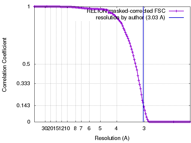
| Method | single particle reconstruction / cryo EM / Resolution: 3.03 Å | |||||||||
 Authors Authors | Brown A / Rathore S | |||||||||
 Citation Citation | Journal: Nat Struct Mol Biol / Year: 2017 Title: Structures of the human mitochondrial ribosome in native states of assembly. Authors: Alan Brown / Sorbhi Rathore / Dari Kimanius / Shintaro Aibara / Xiao-Chen Bai / Joanna Rorbach / Alexey Amunts / V Ramakrishnan /   Abstract: Mammalian mitochondrial ribosomes (mitoribosomes) have less rRNA content and 36 additional proteins compared with the evolutionarily related bacterial ribosome. These differences make the assembly of ...Mammalian mitochondrial ribosomes (mitoribosomes) have less rRNA content and 36 additional proteins compared with the evolutionarily related bacterial ribosome. These differences make the assembly of mitoribosomes more complex than the assembly of bacterial ribosomes, but the molecular details of mitoribosomal biogenesis remain elusive. Here, we report the structures of two late-stage assembly intermediates of the human mitoribosomal large subunit (mt-LSU) isolated from a native pool within a human cell line and solved by cryo-EM to ∼3-Å resolution. Comparison of the structures reveals insights into the timing of rRNA folding and protein incorporation during the final steps of ribosomal maturation and the evolutionary adaptations that are required to preserve biogenesis after the structural diversification of mitoribosomes. Furthermore, the structures redefine the ribosome silencing factor (RsfS) family as multifunctional biogenesis factors and identify two new assembly factors (L0R8F8 and mt-ACP) not previously implicated in mitoribosomal biogenesis. | |||||||||
| History |
|
- Structure visualization
| Movie |
Movie viewer |
|---|---|
| Structure viewer | EM map: SurfViewMolmilJmol/JSmol |
| Supplemental images |
- Downloads & links
-EMDB archive
| Map data | emd_3843.map.gz | 13.7 MB | EMDB map data format | |
|---|---|---|---|---|
| Header (meta data) | emd-3843-v30.xmlemd-3843.xml | 85.5 KB 85.5 KB | Display Display | EMDB header |
| FSC (resolution estimation) | emd_3843_fsc.xml | 12.1 KB | Display | FSC data file |
| Images |  emd_3843.png emd_3843.png | 266.3 KB | ||
| Masks | emd_3843_msk_1.map | 166.4 MB | Mask map | |
| Filedesc metadata | emd-3843.cif.gz | 17.8 KB | ||
| Others | emd_3843_half_map_1.map.gzemd_3843_half_map_2.map.gz | 130.6 MB 130.5 MB | ||
| Archive directory |  http://ftp.pdbj.org/pub/emdb/structures/EMD-3843ftp://ftp.pdbj.org/pub/emdb/structures/EMD-3843 http://ftp.pdbj.org/pub/emdb/structures/EMD-3843ftp://ftp.pdbj.org/pub/emdb/structures/EMD-3843 | HTTPS FTP |
-Related structure data
| Related structure data | 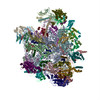 5oomMC 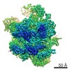 3842C 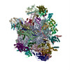 5oolC M: atomic model generated by this map C: citing same article ( |
|---|---|
| Similar structure data |


-Links
| EMDB pages | EMDB (EBI/PDBe) / EMDataResource |
|---|---|
| Related items in Molecule of the Month |


-Map
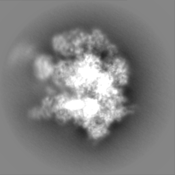
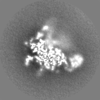
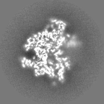
| File | Download / File: emd_3843.map.gz / Format: CCP4 / Size: 166.4 MB / Type: IMAGE STORED AS FLOATING POINT NUMBER (4 BYTES) | ||||||||||||||||||||||||||||||||||||||||||||||||||||||||||||
|---|---|---|---|---|---|---|---|---|---|---|---|---|---|---|---|---|---|---|---|---|---|---|---|---|---|---|---|---|---|---|---|---|---|---|---|---|---|---|---|---|---|---|---|---|---|---|---|---|---|---|---|---|---|---|---|---|---|---|---|---|---|
| Projections & slices | Image control
Images are generated by Spider. | ||||||||||||||||||||||||||||||||||||||||||||||||||||||||||||
| Voxel size | X=Y=Z: 1.06 Å | ||||||||||||||||||||||||||||||||||||||||||||||||||||||||||||
| Density |
| ||||||||||||||||||||||||||||||||||||||||||||||||||||||||||||
| Symmetry | Space group: 1 | ||||||||||||||||||||||||||||||||||||||||||||||||||||||||||||
| Details | EMDB XML:
CCP4 map header:
| ||||||||||||||||||||||||||||||||||||||||||||||||||||||||||||
 Z (Sec.)
 Y (Row.)
 X (Col.)


-Supplemental data
-Mask #1
| File | emd_3843_msk_1.map | ||||||||||||
|---|---|---|---|---|---|---|---|---|---|---|---|---|---|
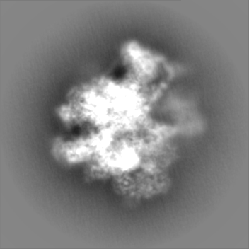
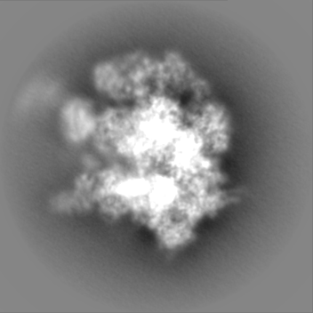
| Projections & Slices |
| ||||||||||||
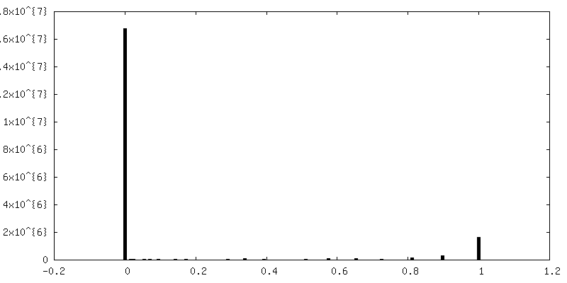
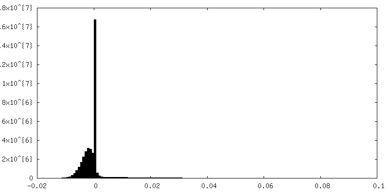
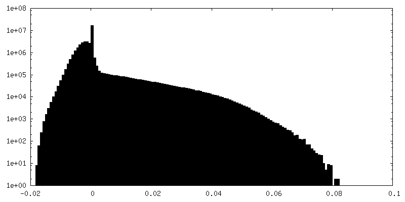
| Density Histograms |

-Half map: #1
| File | emd_3843_half_map_1.map | ||||||||||||
|---|---|---|---|---|---|---|---|---|---|---|---|---|---|
| Projections & Slices |
| ||||||||||||
| Density Histograms |


-Half map: #2
| File | emd_3843_half_map_2.map | ||||||||||||
|---|---|---|---|---|---|---|---|---|---|---|---|---|---|
| Projections & Slices |
| ||||||||||||
| Density Histograms |


- Sample components
+Entire : Mitochondrial ribosome large subunit with assembly factor
+Supramolecule #1: Mitochondrial ribosome large subunit with assembly factor
+Macromolecule #1: 16S ribosomal RNA
+Macromolecule #2: mitochondrial tRNAVal
+Macromolecule #3: 39S ribosomal protein L2, mitochondrial
+Macromolecule #4: 39S ribosomal protein L3, mitochondrial
+Macromolecule #5: 39S ribosomal protein L4, mitochondrial
+Macromolecule #6: 39S ribosomal protein L9, mitochondrial
+Macromolecule #7: 39S ribosomal protein L10, mitochondrial
+Macromolecule #8: 39S ribosomal protein L11, mitochondrial
+Macromolecule #9: 39S ribosomal protein L13, mitochondrial
+Macromolecule #10: 39S ribosomal protein L14, mitochondrial
+Macromolecule #11: 39S ribosomal protein L15, mitochondrial
+Macromolecule #12: 39S ribosomal protein L16, mitochondrial
+Macromolecule #13: 39S ribosomal protein L17, mitochondrial
+Macromolecule #14: 39S ribosomal protein L18, mitochondrial
+Macromolecule #15: 39S ribosomal protein L19, mitochondrial
+Macromolecule #16: 39S ribosomal protein L20, mitochondrial
+Macromolecule #17: 39S ribosomal protein L21, mitochondrial
+Macromolecule #18: 39S ribosomal protein L22, mitochondrial
+Macromolecule #19: 39S ribosomal protein L23, mitochondrial
+Macromolecule #20: 39S ribosomal protein L24, mitochondrial
+Macromolecule #21: 39S ribosomal protein L27, mitochondrial
+Macromolecule #22: 39S ribosomal protein L28, mitochondrial
+Macromolecule #23: 39S ribosomal protein L47, mitochondrial
+Macromolecule #24: 39S ribosomal protein L30, mitochondrial
+Macromolecule #25: 39S ribosomal protein L32, mitochondrial
+Macromolecule #26: 39S ribosomal protein L33, mitochondrial
+Macromolecule #27: 39S ribosomal protein L34, mitochondrial
+Macromolecule #28: 39S ribosomal protein L35, mitochondrial
+Macromolecule #29: 39S ribosomal protein L37, mitochondrial
+Macromolecule #30: 39S ribosomal protein L38, mitochondrial
+Macromolecule #31: 39S ribosomal protein L39, mitochondrial
+Macromolecule #32: 39S ribosomal protein L40, mitochondrial
+Macromolecule #33: 39S ribosomal protein L41, mitochondrial
+Macromolecule #34: 39S ribosomal protein L42, mitochondrial
+Macromolecule #35: 39S ribosomal protein L43, mitochondrial
+Macromolecule #36: 39S ribosomal protein L44, mitochondrial
+Macromolecule #37: 39S ribosomal protein L45, mitochondrial
+Macromolecule #38: 39S ribosomal protein L46, mitochondrial
+Macromolecule #39: 39S ribosomal protein L48, mitochondrial
+Macromolecule #40: 39S ribosomal protein L49, mitochondrial
+Macromolecule #41: 39S ribosomal protein L50, mitochondrial
+Macromolecule #42: 39S ribosomal protein L51, mitochondrial
+Macromolecule #43: 39S ribosomal protein L52, mitochondrial
+Macromolecule #44: 39S ribosomal protein L53, mitochondrial
+Macromolecule #45: 39S ribosomal protein L54, mitochondrial
+Macromolecule #46: 39S ribosomal protein L55, mitochondrial
+Macromolecule #47: Ribosomal protein 63, mitochondrial
+Macromolecule #48: Peptidyl-tRNA hydrolase ICT1, mitochondrial
+Macromolecule #49: Growth arrest and DNA damage-inducible proteins-interacting protein 1
+Macromolecule #50: 39S ribosomal protein S18a, mitochondrial
+Macromolecule #51: 39S ribosomal protein S30, mitochondrial
+Macromolecule #52: Unknown protein or protein extension
+Macromolecule #53: Mitochondrial assembly of ribosomal large subunit protein 1
+Macromolecule #54: MIEF1 upstream open reading frame protein
+Macromolecule #55: Acyl carrier protein, mitochondrial
+Macromolecule #56: MAGNESIUM ION
+Macromolecule #57: ZINC ION
+Macromolecule #58: 4'-PHOSPHOPANTETHEINE
-Experimental details
-Structure determination
| Method | cryo EM |
|---|---|
 Processing Processing | single particle reconstruction |
| Aggregation state | particle |
-Sample preparation
| Buffer | pH: 7.45 Component:
Details: 2 mM Synercid (Santa Cruz Biotechnology, Inc) was added to reduce preferential orientation. | |||||||||||||||
|---|---|---|---|---|---|---|---|---|---|---|---|---|---|---|---|---|
| Grid | Model: Quantifoil R2/2 / Material: COPPER / Mesh: 400 / Support film - Material: CARBON / Support film - topology: CONTINUOUS / Support film - Film thickness: 3 / Pretreatment - Type: GLOW DISCHARGE | |||||||||||||||
| Vitrification | Cryogen name: ETHANE / Chamber humidity: 100 % / Chamber temperature: 277 K / Instrument: FEI VITROBOT MARK IV / Details: Sample incubated for 30 s prior to freezing.. | |||||||||||||||
| Details | Sample concentration = 100 nM |
- Electron microscopy
| Microscope | FEI TITAN KRIOS |
|---|---|
| Image recording | Film or detector model: FEI FALCON II (4k x 4k) / Detector mode: INTEGRATING / Digitization - Frames/image: 1-25 / Average exposure time: 1.5 sec. / Average electron dose: 1.56 e/Å2 |
| Electron beam | Acceleration voltage: 300 kV / Electron source:  FIELD EMISSION GUN FIELD EMISSION GUN |
| Electron optics | Calibrated magnification: 130841 / Illumination mode: FLOOD BEAM / Imaging mode: BRIGHT FIELD |
| Sample stage | Specimen holder model: FEI TITAN KRIOS AUTOGRID HOLDER / Cooling holder cryogen: NITROGEN |
| Experimental equipment |  Model: Titan Krios / Image courtesy: FEI Company |
+Image processing
-Atomic model buiding 1
| Initial model | PDB ID: Chain - Source name: PDB / Chain - Initial model type: experimental model |
|---|---|
| Refinement | Space: REAL / Protocol: OTHER / Overall B value: 85 / Target criteria: Cross-correlation coefficient |
| Output model | PDB-5oom: |