 Movie
Movie Controller

[English] 日本語
 Yorodumi
- EMDB-2879: The structure of the human mitochondrial ribosome (large subunit,... -
+ Open data

- Basic information
| Entry | Database: EMDB / ID: EMD-2879 | |||||||||
|---|---|---|---|---|---|---|---|---|---|---|
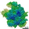
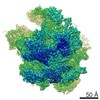
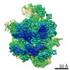
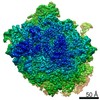
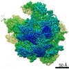
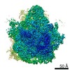
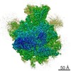
| Title | The structure of the human mitochondrial ribosome (large subunit, masked) | |||||||||
 Map data Map data | Large subunit of the human mitochondrial ribosome (masked). | |||||||||
 Sample Sample |
| |||||||||
 Keywords Keywords | Mitochondrial ribosome / 55S | |||||||||
| Biological species |  Homo sapiens (human) Homo sapiens (human) | |||||||||
| Method | single particle reconstruction / cryo EM / Resolution: 3.27 Å | |||||||||
 Authors Authors | Amunts A / Brown A / Toots J / Scheres SH / Ramakrishnan V | |||||||||
 Citation Citation | Journal: Science / Year: 2015 Title: Ribosome. The structure of the human mitochondrial ribosome. Authors: Alexey Amunts / Alan Brown / Jaan Toots / Sjors H W Scheres / V Ramakrishnan /  Abstract: The highly divergent ribosomes of human mitochondria (mitoribosomes) synthesize 13 essential proteins of oxidative phosphorylation complexes. We have determined the structure of the intact ...The highly divergent ribosomes of human mitochondria (mitoribosomes) synthesize 13 essential proteins of oxidative phosphorylation complexes. We have determined the structure of the intact mitoribosome to 3.5 angstrom resolution by means of single-particle electron cryogenic microscopy. It reveals 80 extensively interconnected proteins, 36 of which are specific to mitochondria, and three ribosomal RNA molecules. The head domain of the small subunit, particularly the messenger (mRNA) channel, is highly remodeled. Many intersubunit bridges are specific to the mitoribosome, which adopts conformations involving ratcheting or rolling of the small subunit that are distinct from those seen in bacteria or eukaryotes. An intrinsic guanosine triphosphatase mediates a contact between the head and central protuberance. The structure provides a reference for analysis of mutations that cause severe pathologies and for future drug design. | |||||||||
| History |
|
- Structure visualization
| Movie |
 Movie viewer Movie viewer |
|---|---|
| Structure viewer | EM map: SurfViewMolmilJmol/JSmol |
| Supplemental images |

- Downloads & links
-EMDB archive
| Map data | emd_2879.map.gz | 8.9 MB | EMDB map data format | |
|---|---|---|---|---|
| Header (meta data) | emd-2879-v30.xmlemd-2879.xml | 15.9 KB 15.9 KB | Display Display | EMDB header |
| FSC (resolution estimation) | emd_2879_fsc.xml | 11.1 KB | Display | FSC data file |
| Images | 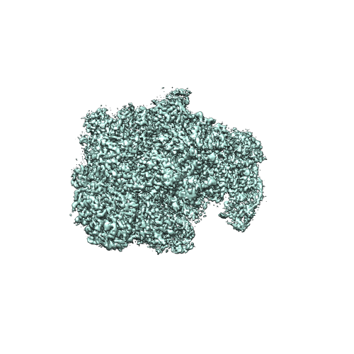 EMD-2879.png EMD-2879.png | 132.1 KB | ||
| Others | emd_2879_half_map_1.map.gzemd_2879_half_map_2.map.gz | 109.6 MB 108.9 MB | ||
| Archive directory |  http://ftp.pdbj.org/pub/emdb/structures/EMD-2879ftp://ftp.pdbj.org/pub/emdb/structures/EMD-2879 http://ftp.pdbj.org/pub/emdb/structures/EMD-2879ftp://ftp.pdbj.org/pub/emdb/structures/EMD-2879 | HTTPS FTP |
-Related structure data
| Related structure data |  2876C  2877C  2878C 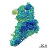 2880C 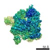 2881C 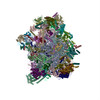 3j9mC C: citing same article ( |
|---|---|
| Similar structure data |


-Links
| EMDB pages | EMDB (EBI/PDBe) / EMDataResource |
|---|---|
| Related items in Molecule of the Month |

-Map
| File | Download / File: emd_2879.map.gz / Format: CCP4 / Size: 122.1 MB / Type: IMAGE STORED AS FLOATING POINT NUMBER (4 BYTES) | ||||||||||||||||||||||||||||||||||||||||||||||||||||||||||||
|---|---|---|---|---|---|---|---|---|---|---|---|---|---|---|---|---|---|---|---|---|---|---|---|---|---|---|---|---|---|---|---|---|---|---|---|---|---|---|---|---|---|---|---|---|---|---|---|---|---|---|---|---|---|---|---|---|---|---|---|---|---|
| Annotation | Large subunit of the human mitochondrial ribosome (masked). | ||||||||||||||||||||||||||||||||||||||||||||||||||||||||||||
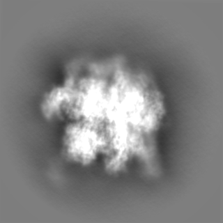
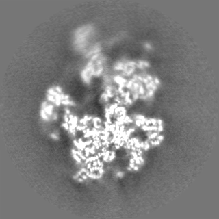
| Projections & slices | Image control
Images are generated by Spider. | ||||||||||||||||||||||||||||||||||||||||||||||||||||||||||||
| Voxel size | X=Y=Z: 1.34 Å | ||||||||||||||||||||||||||||||||||||||||||||||||||||||||||||
| Density |
| ||||||||||||||||||||||||||||||||||||||||||||||||||||||||||||
| Symmetry | Space group: 1 | ||||||||||||||||||||||||||||||||||||||||||||||||||||||||||||
| Details | EMDB XML:
CCP4 map header:
| ||||||||||||||||||||||||||||||||||||||||||||||||||||||||||||
 Z (Sec.)
 Y (Row.)
 X (Col.)


-Supplemental data
-Supplemental map: emd 2879 half map 1.map
| File | emd_2879_half_map_1.map | ||||||||||||
|---|---|---|---|---|---|---|---|---|---|---|---|---|---|
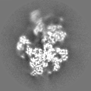
| Projections & Slices |
| ||||||||||||
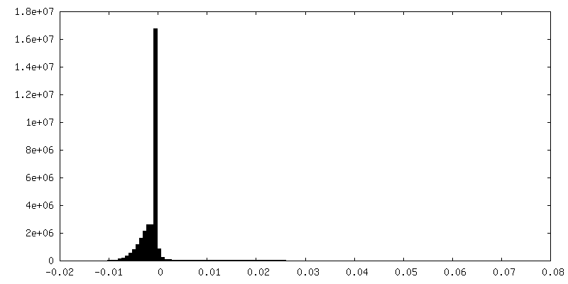
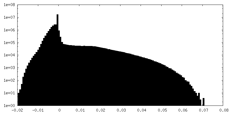
| Density Histograms |


-Supplemental map: emd 2879 half map 2.map
| File | emd_2879_half_map_2.map | ||||||||||||
|---|---|---|---|---|---|---|---|---|---|---|---|---|---|
| Projections & Slices |
| ||||||||||||
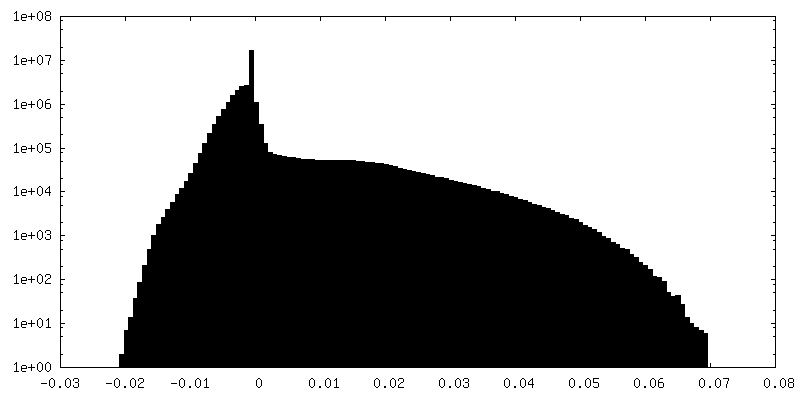
| Density Histograms |


- Sample components
-Entire : Human mitochondrial ribosome
| Entire | Name: Human mitochondrial ribosome |
|---|---|
| Components |
|
-Supramolecule #1000: Human mitochondrial ribosome
| Supramolecule | Name: Human mitochondrial ribosome / type: sample / ID: 1000 / Details: The sample was monodisperse / Oligomeric state: Multimer / Number unique components: 1 |
|---|---|
| Molecular weight | Experimental: 1.7 MDa / Theoretical: 1.7 MDa / Method: Sedimentation |
-Supramolecule #1: Large subunit of the human mitochondrial ribosome
| Supramolecule | Name: Large subunit of the human mitochondrial ribosome / type: complex / ID: 1 / Name.synonym: 39S subunit / Recombinant expression: No / Ribosome-details: ribosome-eukaryote: ALL |
|---|---|
| Source (natural) | Organism: Homo sapiens (human) / synonym: human / Tissue: kidney / Cell: HEK 293 / Organelle: mitochondria / Location in cell: Inner mitochondrial membrane |
| Molecular weight | Experimental: 1.7 MDa / Theoretical: 1.7 MDa |
-Experimental details
-Structure determination
| Method | cryo EM |
|---|---|
 Processing Processing | single particle reconstruction |
| Aggregation state | particle |
-Sample preparation
| Concentration | 0.23 mg/mL |
|---|---|
| Buffer | pH: 7.45 Details: 20 mM Hepes-KOH pH 7.45, 100 mM KCl, 20 mM MgOAc, 2 mM DTT |
| Grid | Details: 30 s on glow-discharged holey carbon grids (Quantifoil R2/2), onto which a home-made continuous carbon film |
| Vitrification | Cryogen name: ETHANE / Chamber humidity: 100 % / Chamber temperature: 90 K / Instrument: FEI VITROBOT MARK II / Method: Blot 2.5 seconds before plunging |
- Electron microscopy #1
| Microscopy ID | 1 |
|---|---|
| Microscope | FEI TITAN KRIOS |
| Temperature | Min: 80 K / Max: 90 K / Average: 85 K |
| Alignment procedure | Legacy - Astigmatism: Objective lens astigmatism was corrected at 59,000 times magnification |
| Specialist optics | Energy filter - Name: FEI |
| Date | Apr 3, 2014 |
| Image recording | Category: CCD / Film or detector model: FEI FALCON II (4k x 4k) / Number real images: 7526 / Average electron dose: 25 e/Å2 Details: Every image is the average of 17 frames recorded by the direct electron detector |
| Electron beam | Acceleration voltage: 300 kV / Electron source:  FIELD EMISSION GUN FIELD EMISSION GUN |
| Electron optics | Calibrated magnification: 104478 / Illumination mode: FLOOD BEAM / Imaging mode: BRIGHT FIELD / Cs: 2.7 mm / Nominal defocus max: 3.5 µm / Nominal defocus min: 1.5 µm / Nominal magnification: 59000 |
| Sample stage | Specimen holder model: FEI TITAN KRIOS AUTOGRID HOLDER |
| Experimental equipment |  Model: Titan Krios / Image courtesy: FEI Company |
-Electron microscopy #2
| Microscopy ID | 2 |
|---|---|
| Microscope | FEI TITAN KRIOS |
| Temperature | Min: 80 K / Max: 90 K / Average: 85 K |
| Alignment procedure | Legacy - Astigmatism: Objective lens astigmatism was corrected at 59,000 times magnification |
| Specialist optics | Energy filter - Name: FEI |
| Date | Apr 11, 2014 |
| Image recording | Category: CCD / Film or detector model: FEI FALCON II (4k x 4k) / Number real images: 7526 / Average electron dose: 25 e/Å2 Details: Every image is the average of 17 frames recorded by the direct electron detector |
| Electron beam | Acceleration voltage: 300 kV / Electron source: FIELD EMISSION GUN |
| Electron optics | Calibrated magnification: 104478 / Illumination mode: FLOOD BEAM / Imaging mode: BRIGHT FIELD / Cs: 2.7 mm / Nominal defocus max: 3.5 µm / Nominal defocus min: 1.5 µm / Nominal magnification: 59000 |
| Sample stage | Specimen holder model: FEI TITAN KRIOS AUTOGRID HOLDER |
| Experimental equipment | Model: Titan Krios / Image courtesy: FEI Company |
-Electron microscopy #3
| Microscopy ID | 3 |
|---|---|
| Microscope | FEI TITAN KRIOS |
| Temperature | Min: 80 K / Max: 90 K / Average: 85 K |
| Alignment procedure | Legacy - Astigmatism: Objective lens astigmatism was corrected at 59,000 times magnification |
| Specialist optics | Energy filter - Name: FEI |
| Date | May 10, 2014 |
| Image recording | Category: CCD / Film or detector model: FEI FALCON II (4k x 4k) / Number real images: 7526 / Average electron dose: 25 e/Å2 Details: Every image is the average of 17 frames recorded by the direct electron detector |
| Electron beam | Acceleration voltage: 300 kV / Electron source: FIELD EMISSION GUN |
| Electron optics | Calibrated magnification: 104478 / Illumination mode: FLOOD BEAM / Imaging mode: BRIGHT FIELD / Cs: 2.7 mm / Nominal defocus max: 3.5 µm / Nominal defocus min: 1.5 µm / Nominal magnification: 59000 |
| Sample stage | Specimen holder model: FEI TITAN KRIOS AUTOGRID HOLDER |
| Experimental equipment | Model: Titan Krios / Image courtesy: FEI Company |
-Electron microscopy #4
| Microscopy ID | 4 |
|---|---|
| Microscope | FEI TITAN KRIOS |
| Temperature | Min: 80 K / Max: 90 K / Average: 85 K |
| Alignment procedure | Legacy - Astigmatism: Objective lens astigmatism was corrected at 59,000 times magnification |
| Specialist optics | Energy filter - Name: FEI |
| Date | May 30, 2014 |
| Image recording | Category: CCD / Film or detector model: FEI FALCON II (4k x 4k) / Number real images: 7526 / Average electron dose: 25 e/Å2 Details: Every image is the average of 17 frames recorded by the direct electron detector |
| Electron beam | Acceleration voltage: 300 kV / Electron source: FIELD EMISSION GUN |
| Electron optics | Calibrated magnification: 104478 / Illumination mode: FLOOD BEAM / Imaging mode: BRIGHT FIELD / Cs: 2.7 mm / Nominal defocus max: 3.5 µm / Nominal defocus min: 1.5 µm / Nominal magnification: 59000 |
| Sample stage | Specimen holder model: FEI TITAN KRIOS AUTOGRID HOLDER |
| Experimental equipment | Model: Titan Krios / Image courtesy: FEI Company |
-Image processing
| Details | Data were processed in Relion to compensate for beam-induced movement. |
|---|---|
| CTF correction | Details: Each particle |
| Final reconstruction | Applied symmetry - Point group: C1 (asymmetric) / Resolution.type: BY AUTHOR / Resolution: 3.27 Å / Resolution method: OTHER / Software - Name: CTFFIND3, RELION / Number images used: 286106 |
| FSC plot (resolution estimation) |  |
