 Movie
Movie Controller

[English] 日本語
 Yorodumi
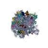
- EMDB-3531: Cryo-EM reconstruction of the large subunit of the chloroplast ri... -
+ Open data

- Basic information
| Entry | Database: EMDB / ID: EMD-3531 | |||||||||
|---|---|---|---|---|---|---|---|---|---|---|
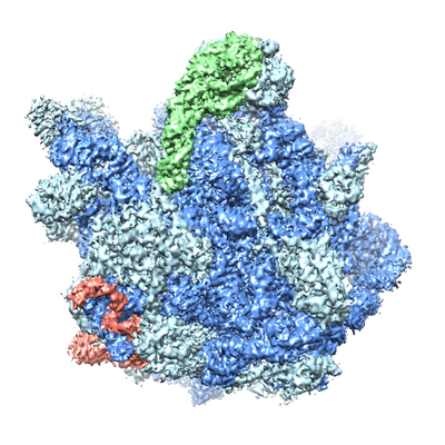
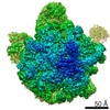
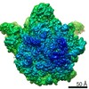
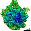
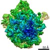
| Title | Cryo-EM reconstruction of the large subunit of the chloroplast ribosome | |||||||||
 Map data Map data | Cryo-EM reconstruction of the 50S large subunit of the chloroplast ribosome | |||||||||
 Sample Sample |
| |||||||||
 Keywords Keywords | chloroplast / translation / ribosome / cryo-EM | |||||||||
| Function / homology |  Function and homology information Function and homology informationplastid translation / chloroplast envelope / mitochondrial large ribosomal subunit / mitochondrial translation / chloroplast thylakoid membrane / chloroplast / DNA-templated transcription termination / large ribosomal subunit / transferase activity / 5S rRNA binding ...plastid translation / chloroplast envelope / mitochondrial large ribosomal subunit / mitochondrial translation / chloroplast thylakoid membrane / chloroplast / DNA-templated transcription termination / large ribosomal subunit / transferase activity / 5S rRNA binding / ribosomal large subunit assembly / large ribosomal subunit rRNA binding / cytosolic large ribosomal subunit / negative regulation of translation / rRNA binding / structural constituent of ribosome / ribosome / translation / ribonucleoprotein complex / mRNA binding / mitochondrion / RNA binding Similarity search - Function | |||||||||
| Biological species |  Spinacia oleracea (spinach) Spinacia oleracea (spinach) | |||||||||
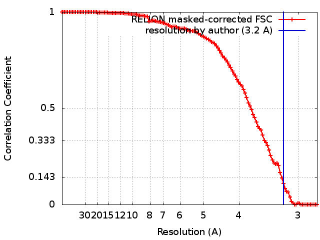
| Method | single particle reconstruction / cryo EM / Resolution: 3.2 Å | |||||||||
 Authors Authors | Bieri P / Leibundgut M | |||||||||
 Citation Citation | Journal: EMBO J / Year: 2017 Title: The complete structure of the chloroplast 70S ribosome in complex with translation factor pY. Authors: Philipp Bieri / Marc Leibundgut / Martin Saurer / Daniel Boehringer / Nenad Ban /  Abstract: Chloroplasts are cellular organelles of plants and algae that are responsible for energy conversion and carbon fixation by the photosynthetic reaction. As a consequence of their endosymbiotic origin, ...Chloroplasts are cellular organelles of plants and algae that are responsible for energy conversion and carbon fixation by the photosynthetic reaction. As a consequence of their endosymbiotic origin, they still contain their own genome and the machinery for protein biosynthesis. Here, we present the atomic structure of the chloroplast 70S ribosome prepared from spinach leaves and resolved by cryo-EM at 3.4 Å resolution. The complete structure reveals the features of the 4.5S rRNA, which probably evolved by the fragmentation of the 23S rRNA, and all five plastid-specific ribosomal proteins. These proteins, required for proper assembly and function of the chloroplast translation machinery, bind and stabilize rRNA including regions that only exist in the chloroplast ribosome. Furthermore, the structure reveals plastid-specific extensions of ribosomal proteins that extensively remodel the mRNA entry and exit site on the small subunit as well as the polypeptide tunnel exit and the putative binding site of the signal recognition particle on the large subunit. The translation factor pY, involved in light- and temperature-dependent control of protein synthesis, is bound to the mRNA channel of the small subunit and interacts with 16S rRNA nucleotides at the A-site and P-site, where it protects the decoding centre and inhibits translation by preventing tRNA binding. The small subunit is locked by pY in a non-rotated state, in which the intersubunit bridges to the large subunit are stabilized. | |||||||||
| History |
|
- Structure visualization
| Movie |
Movie viewer |
|---|---|
| Structure viewer | EM map: SurfViewMolmilJmol/JSmol |
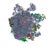
| Supplemental images |
- Downloads & links
-EMDB archive
| Map data | emd_3531.map.gz | 10.8 MB | EMDB map data format | |
|---|---|---|---|---|
| Header (meta data) | emd-3531-v30.xmlemd-3531.xml | 51.2 KB 51.2 KB | Display Display | EMDB header |
| FSC (resolution estimation) | emd_3531_fsc.xml | 11.2 KB | Display | FSC data file |
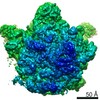
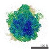
| Images |  emd_3531.png emd_3531.png | 207 KB | ||
| Filedesc metadata | emd-3531.cif.gz | 11.9 KB | ||
| Archive directory |  http://ftp.pdbj.org/pub/emdb/structures/EMD-3531ftp://ftp.pdbj.org/pub/emdb/structures/EMD-3531 http://ftp.pdbj.org/pub/emdb/structures/EMD-3531ftp://ftp.pdbj.org/pub/emdb/structures/EMD-3531 | HTTPS FTP |
-Validation report
| Summary document | emd_3531_validation.pdf.gz | 265.8 KB | Display | EMDB validaton report |
|---|---|---|---|---|
| Full document | emd_3531_full_validation.pdf.gz | 264.9 KB | Display | |
| Data in XML | emd_3531_validation.xml.gz | 12.2 KB | Display | |
| Arichive directory | https://ftp.pdbj.org/pub/emdb/validation_reports/EMD-3531ftp://ftp.pdbj.org/pub/emdb/validation_reports/EMD-3531 | HTTPS FTP |
-Related structure data
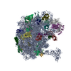
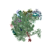
| Related structure data |  5mmiMC  3532C  3533C 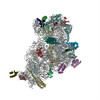 5mmjC 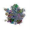 5mmmC M: atomic model generated by this map C: citing same article ( |
|---|---|
| Similar structure data |

-Links
| EMDB pages | EMDB (EBI/PDBe) / EMDataResource |
|---|---|
| Related items in Molecule of the Month |


-Map
| File | Download / File: emd_3531.map.gz / Format: CCP4 / Size: 125 MB / Type: IMAGE STORED AS FLOATING POINT NUMBER (4 BYTES) | ||||||||||||||||||||||||||||||||||||||||||||||||||||||||||||||||||||
|---|---|---|---|---|---|---|---|---|---|---|---|---|---|---|---|---|---|---|---|---|---|---|---|---|---|---|---|---|---|---|---|---|---|---|---|---|---|---|---|---|---|---|---|---|---|---|---|---|---|---|---|---|---|---|---|---|---|---|---|---|---|---|---|---|---|---|---|---|---|
| Annotation | Cryo-EM reconstruction of the 50S large subunit of the chloroplast ribosome | ||||||||||||||||||||||||||||||||||||||||||||||||||||||||||||||||||||
| Projections & slices | Image control
Images are generated by Spider. | ||||||||||||||||||||||||||||||||||||||||||||||||||||||||||||||||||||
| Voxel size | X=Y=Z: 1.39 Å | ||||||||||||||||||||||||||||||||||||||||||||||||||||||||||||||||||||
| Density |
| ||||||||||||||||||||||||||||||||||||||||||||||||||||||||||||||||||||
| Symmetry | Space group: 1 | ||||||||||||||||||||||||||||||||||||||||||||||||||||||||||||||||||||
| Details | EMDB XML:
CCP4 map header:
| ||||||||||||||||||||||||||||||||||||||||||||||||||||||||||||||||||||
 Z (Sec.)
 Y (Row.)
 X (Col.)


-Supplemental data
- Sample components
+Entire : Chloroplast 70S ribosome
+Supramolecule #1: Chloroplast 70S ribosome
+Macromolecule #1: 50S ribosomal protein L31
+Macromolecule #2: 50S ribosomal protein L32, chloroplastic
+Macromolecule #3: 50S ribosomal protein L33, chloroplastic
+Macromolecule #4: 50S ribosomal protein L34, chloroplastic
+Macromolecule #5: 50S ribosomal protein L35, chloroplastic
+Macromolecule #6: 50S ribosomal protein L36, chloroplastic
+Macromolecule #7: plastid ribosomal protein cL37, PSRP5
+Macromolecule #8: 50S ribosomal protein 6, chloroplastic
+Macromolecule #11: 50S ribosomal protein L2, chloroplastic
+Macromolecule #12: plastid ribosomal protein uL3c
+Macromolecule #13: plastid ribosomal protein uL4c
+Macromolecule #14: plastid ribosomal protein uL5c
+Macromolecule #15: plastid ribosomal protein uL6c
+Macromolecule #16: plastid ribosomal protein bL9c
+Macromolecule #17: plastid ribosomal protein uL10c
+Macromolecule #18: 50S ribosomal protein L11, chloroplastic
+Macromolecule #19: 50S ribosomal protein L13, chloroplastic
+Macromolecule #20: 50S ribosomal protein L14, chloroplastic
+Macromolecule #21: plastid ribosomal protein uL15c
+Macromolecule #22: 50S ribosomal protein L16, chloroplastic
+Macromolecule #23: plastid ribosomal protein bL17c
+Macromolecule #24: plastid ribosomal protein uL18c
+Macromolecule #25: 50S ribosomal protein L19, chloroplastic
+Macromolecule #26: 50S ribosomal protein L20, chloroplastic
+Macromolecule #27: 50S ribosomal protein L21, chloroplastic
+Macromolecule #28: 50S ribosomal protein L22, chloroplastic
+Macromolecule #29: 50S ribosomal protein L23, chloroplastic
+Macromolecule #30: plastid ribosomal protein uL24c
+Macromolecule #32: plastid ribosomal protein bL27c
+Macromolecule #33: plastid ribosomal protein bL28c
+Macromolecule #34: plastid ribosomal protein uL29c
+Macromolecule #9: 23S ribosomal RNA
+Macromolecule #10: 5S ribosomal RNA
+Macromolecule #31: 4.5S ribosomal RNA
+Macromolecule #35: E-site tRNA
+Macromolecule #36: ZINC ION
+Macromolecule #37: MAGNESIUM ION
-Experimental details
-Structure determination
| Method | cryo EM |
|---|---|
 Processing Processing | single particle reconstruction |
| Aggregation state | particle |
-Sample preparation
| Concentration | 0.12 mg/mL | |||||||||||||||||||||
|---|---|---|---|---|---|---|---|---|---|---|---|---|---|---|---|---|---|---|---|---|---|---|
| Buffer | pH: 7.6 Component:
| |||||||||||||||||||||
| Grid | Model: Quantifoil R2/2 / Material: COPPER / Support film - Material: CARBON / Support film - topology: CONTINUOUS / Pretreatment - Type: GLOW DISCHARGE | |||||||||||||||||||||
| Vitrification | Cryogen name: ETHANE-PROPANE / Chamber humidity: 100 % / Chamber temperature: 278 K / Instrument: FEI VITROBOT MARK I |
- Electron microscopy
| Microscope | FEI TITAN KRIOS |
|---|---|
| Image recording | Film or detector model: FEI FALCON II (4k x 4k) / Detector mode: INTEGRATING / Digitization - Frames/image: 2-8 / Number real images: 2796 / Average electron dose: 20.0 e/Å2 |
| Electron beam | Acceleration voltage: 300 kV / Electron source:  FIELD EMISSION GUN FIELD EMISSION GUN |
| Electron optics | Illumination mode: FLOOD BEAM / Imaging mode: BRIGHT FIELD / Cs: 2.7 mm / Nominal defocus max: 3.5 µm / Nominal defocus min: 0.8 µm / Nominal magnification: 59000 |
| Sample stage | Specimen holder model: FEI TITAN KRIOS AUTOGRID HOLDER |
| Experimental equipment |  Model: Titan Krios / Image courtesy: FEI Company |
+Image processing
-Atomic model buiding 1
| Refinement | Space: RECIPROCAL / Protocol: OTHER / Overall B value: 56.9 |
|---|---|
| Output model | PDB-5mmi: |
