National Natural Science Foundation of China (NSFC)
China
Citation
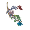
Journal: Cell Discov / Year: 2023 Title: Two assembly modes for SIN3 histone deacetylase complexes. Authors: Chengcheng Wang / Zhouyan Guo / Chen Chu / Yichen Lu / Xiaofeng Zhang / Xiechao Zhan / Abstract: The switch-independent 3 (SIN3)/histone deacetylase (HDAC) complexes play essential roles in regulating chromatin accessibility and gene expression. There are two major types of SIN3/HDAC complexes ...The switch-independent 3 (SIN3)/histone deacetylase (HDAC) complexes play essential roles in regulating chromatin accessibility and gene expression. There are two major types of SIN3/HDAC complexes (named SIN3L and SIN3S) targeting different chromatin regions. Here we present the cryo-electron microscopy structures of the SIN3L and SIN3S complexes from Schizosaccharomyces pombe (S. pombe), revealing two distinct assembly modes. In the structure of SIN3L, each Sin3 isoform (Pst1 and Pst3) interacts with one histone deacetylase Clr6, and one WD40-containing protein Prw1, forming two lobes. These two lobes are bridged by two vertical coiled-coil domains from Sds3/Dep1 and Rxt2/Png2, respectively. In the structure of SIN3S, there is only one lobe organized by another Sin3 isoform Pst2; each of the Cph1 and Cph2 binds to an Eaf3 molecule, providing two modules for histone recognition and binding. Notably, the Pst1 Lobe in SIN3L and the Pst2 Lobe in SIN3S adopt similar conformation with their deacetylase active sites exposed to the space; however, the Pst3 Lobe in SIN3L is in a compact state with its active center buried inside and blocked. Our work reveals two classical organization mechanisms for the SIN3/HDAC complexes to achieve specific targeting and provides a framework for studying the histone deacetylase complexes.
In the structure databanks used in Yorodumi, some data are registered as the other names, "COVID-19 virus" and "2019-nCoV". Here are the details of the virus and the list of structure data.
Jan 31, 2019. EMDB accession codes are about to change! (news from PDBe EMDB page)
EMDB accession codes are about to change! (news from PDBe EMDB page)
The allocation of 4 digits for EMDB accession codes will soon come to an end. Whilst these codes will remain in use, new EMDB accession codes will include an additional digit and will expand incrementally as the available range of codes is exhausted. The current 4-digit format prefixed with “EMD-” (i.e. EMD-XXXX) will advance to a 5-digit format (i.e. EMD-XXXXX), and so on. It is currently estimated that the 4-digit codes will be depleted around Spring 2019, at which point the 5-digit format will come into force.
The EM Navigator/Yorodumi systems omit the EMD- prefix.
Related info.:Q: What is EMD? / ID/Accession-code notation in Yorodumi/EM Navigator
Yorodumi is a browser for structure data from EMDB, PDB, SASBDB, etc.
This page is also the successor to EM Navigator detail page, and also detail information page/front-end page for Omokage search.
The word "yorodu" (or yorozu) is an old Japanese word meaning "ten thousand". "mi" (miru) is to see.
Related info.:EMDB / PDB / SASBDB / Comparison of 3 databanks / Yorodumi Search / Aug 31, 2016. New EM Navigator & Yorodumi / Yorodumi Papers / Jmol/JSmol / Function and homology information / Changes in new EM Navigator and Yorodumi
 Movie
Movie Controller


 Open data
 Basic information
 Map data
 Sample
 Authors
 China, 1 items
  Citation
 Structure visualization
 Downloads & links
 EMDB map data format
 emd_35095.png
 http://ftp.pdbj.org/pub/emdb/structures/EMD-35095

 Sample components
 Processing
 Electron microscopy
 FIELD EMISSION GUN
