 Movie
Movie Controller


+ Open data

- Basic information
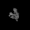
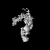
| Entry | Database: PDB / ID: 8i03 | |||||||||||||||||||||||||||||||||
|---|---|---|---|---|---|---|---|---|---|---|---|---|---|---|---|---|---|---|---|---|---|---|---|---|---|---|---|---|---|---|---|---|---|---|
| Title | Cryo-EM structure of the SIN3L complex from S. pombe | |||||||||||||||||||||||||||||||||
 Components Components |
| |||||||||||||||||||||||||||||||||
 Keywords Keywords | DNA BINDING PROTEIN / SIN3 / SIN3L / Pst1 / Pst3 / Clr6 / deacetylase | |||||||||||||||||||||||||||||||||
| Function / homology |  Function and homology information Function and homology informationRegulation of TP53 Activity through Acetylation / PI5P Regulates TP53 Acetylation / SUMOylation of transcription cofactors / HATs acetylate histones / histone H4K16 deacetylase activity, hydrolytic mechanism / histone H4K5 deacetylase activity, hydrolytic mechanism / histone H4K8 deacetylase activity, hydrolytic mechanism / histone H3K14 deacetylase activity, hydrolytic mechanism / Ub-specific processing proteases / HDACs deacetylate histones ...Regulation of TP53 Activity through Acetylation / PI5P Regulates TP53 Acetylation / SUMOylation of transcription cofactors / HATs acetylate histones / histone H4K16 deacetylase activity, hydrolytic mechanism / histone H4K5 deacetylase activity, hydrolytic mechanism / histone H4K8 deacetylase activity, hydrolytic mechanism / histone H3K14 deacetylase activity, hydrolytic mechanism / Ub-specific processing proteases / HDACs deacetylate histones / histone H4K12 deacetylase activity, hydrolytic mechanism / SUMOylation of chromatin organization proteins / H3-H4 histone complex chaperone activity / histone H3K9 deacetylase activity, hydrolytic mechanism / Rpd3L complex / Rpd3L-Expanded complex / Rpd3S complex / pericentric heterochromatin formation / histone deacetylase / protein lysine deacetylase activity / histone deacetylase activity / DNA repair-dependent chromatin remodeling / : / Sin3-type complex / NuA4 histone acetyltransferase complex / pericentric heterochromatin / transcription initiation-coupled chromatin remodeling / epigenetic regulation of gene expression / double-strand break repair via nonhomologous end joining / histone deacetylase binding / transcription corepressor activity / heterochromatin formation / histone binding / chromatin remodeling / cell division / regulation of DNA-templated transcription / regulation of transcription by RNA polymerase II / chromatin / negative regulation of transcription by RNA polymerase II / zinc ion binding / nucleus / cytosol / cytoplasm Similarity search - Function | |||||||||||||||||||||||||||||||||
| Biological species |  | |||||||||||||||||||||||||||||||||
| Method | ELECTRON MICROSCOPY / single particle reconstruction / cryo EM / Resolution: 3.2 Å | |||||||||||||||||||||||||||||||||
 Authors Authors | Wang, C. / Guo, Z. / Zhan, X. | |||||||||||||||||||||||||||||||||
| Funding support |  China, 1items China, 1items
| |||||||||||||||||||||||||||||||||
 Citation Citation | Journal: Cell Discov / Year: 2023 Title: Two assembly modes for SIN3 histone deacetylase complexes. Authors: Chengcheng Wang / Zhouyan Guo / Chen Chu / Yichen Lu / Xiaofeng Zhang / Xiechao Zhan / Abstract: The switch-independent 3 (SIN3)/histone deacetylase (HDAC) complexes play essential roles in regulating chromatin accessibility and gene expression. There are two major types of SIN3/HDAC complexes ...The switch-independent 3 (SIN3)/histone deacetylase (HDAC) complexes play essential roles in regulating chromatin accessibility and gene expression. There are two major types of SIN3/HDAC complexes (named SIN3L and SIN3S) targeting different chromatin regions. Here we present the cryo-electron microscopy structures of the SIN3L and SIN3S complexes from Schizosaccharomyces pombe (S. pombe), revealing two distinct assembly modes. In the structure of SIN3L, each Sin3 isoform (Pst1 and Pst3) interacts with one histone deacetylase Clr6, and one WD40-containing protein Prw1, forming two lobes. These two lobes are bridged by two vertical coiled-coil domains from Sds3/Dep1 and Rxt2/Png2, respectively. In the structure of SIN3S, there is only one lobe organized by another Sin3 isoform Pst2; each of the Cph1 and Cph2 binds to an Eaf3 molecule, providing two modules for histone recognition and binding. Notably, the Pst1 Lobe in SIN3L and the Pst2 Lobe in SIN3S adopt similar conformation with their deacetylase active sites exposed to the space; however, the Pst3 Lobe in SIN3L is in a compact state with its active center buried inside and blocked. Our work reveals two classical organization mechanisms for the SIN3/HDAC complexes to achieve specific targeting and provides a framework for studying the histone deacetylase complexes. | |||||||||||||||||||||||||||||||||
| History |
|
- Structure visualization
| Structure viewer | Molecule: MolmilJmol/JSmol |
|---|
- Downloads & links
-Download
| PDBx/mmCIF format | 8i03.cif.gz | 700 KB | Display | PDBx/mmCIF format |
|---|---|---|---|---|
| PDB format | pdb8i03.ent.gz | 542.4 KB | Display | PDB format |
| PDBx/mmJSON format | 8i03.json.gz | Tree view | PDBx/mmJSON format | |
| Others |  Other downloads Other downloads |
-Validation report
| Arichive directory | https://data.pdbj.org/pub/pdb/validation_reports/i0/8i03ftp://data.pdbj.org/pub/pdb/validation_reports/i0/8i03 | HTTPS FTP |
|---|
-Related structure data
| Related structure data | 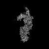 35093MC 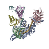 8i02C M: map data used to model this data C: citing same article ( |
|---|---|
| Similar structure data |

-Links
 PDBj
PDBj- Assembly
| Deposited unit | 
|
|---|---|
| 1 |
|
-Components
-Paired amphipathic helix protein ... , 2 types, 2 molecules AB
| #1: Protein | Mass: 171667.125 Da / Num. of mol.: 1 / Source method: isolated from a natural source / Source: (natural) |
|---|---|
| #2: Protein | Mass: 133049.766 Da / Num. of mol.: 1 / Source method: isolated from a natural source / Source: (natural) |
-Protein , 3 types, 5 molecules CDHJK
| #3: Protein | Mass: 46165.844 Da / Num. of mol.: 2 / Source method: isolated from a natural source / Source: (natural) #7: Protein | | Mass: 34925.855 Da / Num. of mol.: 1 / Source method: isolated from a natural source / Source: (natural) #9: Protein | Mass: 48528.926 Da / Num. of mol.: 2 / Source method: isolated from a natural source / Source: (natural) |
|---|
-Transcriptional regulatory protein ... , 4 types, 4 molecules EFGI
| #4: Protein | Mass: 55701.512 Da / Num. of mol.: 1 / Source method: isolated from a natural source / Source: (natural) |
|---|---|
| #5: Protein | Mass: 27429.260 Da / Num. of mol.: 1 / Source method: isolated from a natural source / Source: (natural) |
| #6: Protein | Mass: 30800.586 Da / Num. of mol.: 1 / Source method: isolated from a natural source / Source: (natural) |
| #8: Protein | Mass: 39464.164 Da / Num. of mol.: 1 / Source method: isolated from a natural source / Source: (natural) |
-Non-polymers , 2 types, 6 molecules 

| #10: Chemical |  Mass: 65.409 Da / Num. of mol.: 2 / Source method: obtained synthetically / Formula: Zn Mass: 65.409 Da / Num. of mol.: 2 / Source method: obtained synthetically / Formula: Zn#11: Chemical | ChemComp-K /  Mass: 39.098 Da / Num. of mol.: 4 / Source method: obtained synthetically / Formula: K Mass: 39.098 Da / Num. of mol.: 4 / Source method: obtained synthetically / Formula: K |
|---|
-Details
| Has ligand of interest | N |
|---|---|
| Has protein modification | Y |
-Experimental details
-Experiment
| Experiment | Method: ELECTRON MICROSCOPY |
|---|---|
| EM experiment | Aggregation state: PARTICLE / 3D reconstruction method: single particle reconstruction |
- Sample preparation
| Component | Name: The SIN3S complex / Type: COMPLEX / Entity ID: #1-#5 / Source: NATURAL |
|---|---|
| Source (natural) | Organism: |
| Buffer solution | pH: 7.4 |
| Specimen | Embedding applied: NO / Shadowing applied: NO / Staining applied: NO / Vitrification applied: YES |
| Vitrification | Cryogen name: ETHANE |
- Electron microscopy imaging
| Experimental equipment |  Model: Titan Krios / Image courtesy: FEI Company |
|---|---|
| Microscopy | Model: FEI TITAN KRIOS |
| Electron gun | Electron source:  FIELD EMISSION GUN / Accelerating voltage: 300 kV / Illumination mode: FLOOD BEAM FIELD EMISSION GUN / Accelerating voltage: 300 kV / Illumination mode: FLOOD BEAM |
| Electron lens | Mode: BRIGHT FIELD / Nominal defocus max: 2300 nm / Nominal defocus min: 1800 nm |
| Image recording | Electron dose: 50 e/Å2 / Film or detector model: GATAN K3 (6k x 4k) |
- Processing
| Software | Name: PHENIX / Version: 1.17.1_3660: / Classification: refinement | ||||||||||||||||||||||||
|---|---|---|---|---|---|---|---|---|---|---|---|---|---|---|---|---|---|---|---|---|---|---|---|---|---|
| EM software | Name: PHENIX / Category: model refinement | ||||||||||||||||||||||||
| CTF correction | Type: NONE | ||||||||||||||||||||||||
| 3D reconstruction | Resolution: 3.2 Å / Resolution method: FSC 0.143 CUT-OFF / Num. of particles: 389222 / Symmetry type: POINT | ||||||||||||||||||||||||
| Refinement | Highest resolution: 3.2 Å | ||||||||||||||||||||||||
| Refine LS restraints |
|
