[English] 日本語
Yorodumi
- EMDB-32691: Cryo-EM structure of VWF D1D2 dimer -

+
Open data


ID or keywords:

Loading...

-
Basic information

Entry
Database: EMDB / ID: EMD-32691
TitleCryo-EM structure of VWF D1D2 dimer
Map data
Sample
  • Complex: VWF D1D2 complex
Keywordsblood / VWF / von Willebrand factor / von Willebrand disease / blood coagulation / blood clotting / multimer assembly / VWF assembly / D'D3 domain / D1D2 domain / D'D3 dimer / D1D2 Dimer / VWF Tube / repeating unit
Function / homology
Function and homology information


Defective VWF binding to collagen type I / Enhanced cleavage of VWF variant by ADAMTS13 / Defective VWF cleavage by ADAMTS13 variant / Weibel-Palade body / Defective F8 binding to von Willebrand factor / Enhanced binding of GP1BA variant to VWF multimer:collagen / Defective binding of VWF variant to GPIb:IX:V / hemostasis / platelet alpha granule / Platelet Adhesion to exposed collagen ...Defective VWF binding to collagen type I / Enhanced cleavage of VWF variant by ADAMTS13 / Defective VWF cleavage by ADAMTS13 variant / Weibel-Palade body / Defective F8 binding to von Willebrand factor / Enhanced binding of GP1BA variant to VWF multimer:collagen / Defective binding of VWF variant to GPIb:IX:V / hemostasis / platelet alpha granule / Platelet Adhesion to exposed collagen / positive regulation of intracellular signal transduction / GP1b-IX-V activation signalling / p130Cas linkage to MAPK signaling for integrins / cell-substrate adhesion / Defective F8 cleavage by thrombin / Platelet Aggregation (Plug Formation) / immunoglobulin binding / GRB2:SOS provides linkage to MAPK signaling for Integrins / Integrin cell surface interactions / collagen binding / Intrinsic Pathway of Fibrin Clot Formation / Integrin signaling / extracellular matrix / platelet alpha granule lumen / Signaling by high-kinase activity BRAF mutants / MAP2K and MAPK activation / platelet activation / response to wounding / Signaling by RAF1 mutants / Signaling by moderate kinase activity BRAF mutants / Paradoxical activation of RAF signaling by kinase inactive BRAF / Signaling downstream of RAS mutants / Signaling by BRAF and RAF1 fusions / blood coagulation / integrin binding / Platelet degranulation / protein-folding chaperone binding / collagen-containing extracellular matrix / protease binding / cell adhesion / endoplasmic reticulum / extracellular space / extracellular exosome / extracellular region / identical protein binding
Similarity search - Function
von Willebrand factor, VWA N-terminal domain / Von Willebrand factor / VWA N-terminal / C8 domain / Uncharacterised domain, cysteine-rich / C8 / von Willebrand factor, type D domain / von Willebrand factor type D domain / VWFD domain profile. / von Willebrand factor (vWF) type D domain ...von Willebrand factor, VWA N-terminal domain / Von Willebrand factor / VWA N-terminal / C8 domain / Uncharacterised domain, cysteine-rich / C8 / von Willebrand factor, type D domain / von Willebrand factor type D domain / VWFD domain profile. / von Willebrand factor (vWF) type D domain / C-terminal cystine knot signature. / von Willebrand factor (vWF) type C domain / Trypsin Inhibitor-like, cysteine rich domain / Serine protease inhibitor-like superfamily / Trypsin Inhibitor like cysteine rich domain / C-terminal cystine knot domain profile. / Cystine knot, C-terminal / C-terminal cystine knot-like domain (CTCK) / von Willebrand factor type C domain / VWFC domain signature. / VWFC domain profile. / von Willebrand factor (vWF) type C domain / VWFC domain / von Willebrand factor type A domain / von Willebrand factor (vWF) type A domain / VWFA domain profile. / von Willebrand factor, type A / von Willebrand factor A-like domain superfamily
Similarity search - Domain/homology
von Willebrand factor
Similarity search - Component
Biological speciesHomo sapiens (human)
Methodsingle particle reconstruction / cryo EM / Resolution: 11.4 Å
AuthorsZeng JW / Shu ZM / Zhou AW
Funding support China, 1 items
OrganizationGrant numberCountry
Ministry of Science and Technology (MoST, China)2016YFA0501103 China
Citation
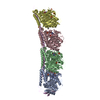
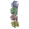
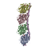
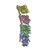
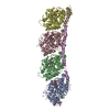
Journal: Blood / Year: 2022
Title: Structural basis of von Willebrand factor multimerization and tubular storage.
Authors: Jianwei Zeng / Zimei Shu / Qian Liang / Jing Zhang / Wenman Wu / Xuefeng Wang / Aiwu Zhou /
Abstract: The von Willebrand factor (VWF) propeptide (domains D1D2) is essential for the assembly of VWF multimers and its tubular storage in Weibel-Palade bodies. However, detailed molecular mechanism ...The von Willebrand factor (VWF) propeptide (domains D1D2) is essential for the assembly of VWF multimers and its tubular storage in Weibel-Palade bodies. However, detailed molecular mechanism underlying this propeptide dependence is unclear. Here, we prepared Weibel-Palade body-like tubules using the N-terminal fragment of VWF and solved the cryo-electron microscopy structures of the tubule at atomic resolution. Detailed structural and biochemical analysis indicate that the propeptide forms a homodimer at acidic pH through the D2:D2 binding interface and then recruits 2 D'D3 domains, forming an intertwined D1D2D'D3 homodimer in essence. Stacking of these homodimers by the intermolecular D1:D2 interfaces brings 2 D3 domains face-to-face and facilitates their disulfide linkages and multimerization of VWF. Sequential stacking of these homodimers leads to a right-hand helical tubule for VWF storage. The clinically identified VWF mutations in the propeptide disrupted different steps of the assembling process, leading to diminished VWF multimers in von Willebrand diseases (VWD). Overall, these results indicate that the propeptide serves as a pH-sensing template for VWF multimerization and tubular storage. This sheds light on delivering normal propeptide as a template to rectify the defects in multimerization of VWD mutants.
History
DepositionJan 24, 2022-
Header (metadata) releaseMar 9, 2022-
Map releaseMar 9, 2022-
UpdateDec 13, 2023-
Current statusDec 13, 2023Processing site: PDBj / Status: Released

-
Structure visualization
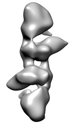
Movie
  • Surface view with section colored by density value
  • Surface level: 0.006
  • Imaged by UCSF Chimera
  • Download
  • Surface view colored by cylindrical radius
  • Surface level: 0.006
  • Imaged by UCSF Chimera
  • Download
Movie viewer
Structure viewerEM map:
SurfViewMolmilJmol/JSmol
Supplemental images

Downloads & links

-
Map

FileDownload / File: emd_32691.map.gz / Format: CCP4 / Size: 8 MB / Type: IMAGE STORED AS FLOATING POINT NUMBER (4 BYTES)
Voxel sizeX=Y=Z: 1.872 Å
Density
Contour LevelBy AUTHOR: 0.006 / Movie #1: 0.006
Minimum - Maximum-0.013845564 - 0.10848707
Average (Standard dev.)0.00019615081 (±0.0029638524)
SymmetrySpace group: 1
Details

EMDB XML:

Map geometry
Axis orderXYZ
Origin000
Dimensions128128128
Spacing128128128
CellA=B=C: 239.616 Å
α=β=γ: 90.0 °

CCP4 map header:

modeImage stored as Reals
Å/pix. X/Y/Z1.8721.8721.872
M x/y/z128128128
origin x/y/z0.0000.0000.000
length x/y/z239.616239.616239.616
α/β/γ90.00090.00090.000
start NX/NY/NZ000
NX/NY/NZ400400400
MAP C/R/S123
start NC/NR/NS000
NC/NR/NS128128128
D min/max/mean-0.0140.1080.000

-
Supplemental data

-
Sample components

-
Entire : VWF D1D2 complex

EntireName: VWF D1D2 complex
Components
  • Complex: VWF D1D2 complex

-
Supramolecule #1: VWF D1D2 complex

SupramoleculeName: VWF D1D2 complex / type: complex / ID: 1 / Parent: 0 / Macromolecule list: #1
Source (natural)Organism: Homo sapiens (human)

-
Experimental details

-
Structure determination

Methodcryo EM
Processingsingle particle reconstruction
Aggregation stateparticle

-
Sample preparation

BufferpH: 7.4
VitrificationCryogen name: ETHANE

-
Electron microscopy

MicroscopeFEI TALOS ARCTICA
Image recordingFilm or detector model: GATAN K2 SUMMIT (4k x 4k) / Average electron dose: 50.0 e/Å2
Electron beamAcceleration voltage: 200 kV / Electron source: FIELD EMISSION GUN
Electron opticsIllumination mode: OTHER / Imaging mode: OTHER / Nominal defocus max: 2.0 µm / Nominal defocus min: 1.0 µm
Experimental equipment
Model: Talos Arctica / Image courtesy: FEI Company

-
Image processing

Startup modelType of model: INSILICO MODEL
Final reconstructionResolution.type: BY AUTHOR / Resolution: 11.4 Å / Resolution method: FSC 0.143 CUT-OFF / Number images used: 199496
Initial angle assignmentType: MAXIMUM LIKELIHOOD
Final angle assignmentType: MAXIMUM LIKELIHOOD

+
About Yorodumi

-
News

-
Feb 9, 2022. New format data for meta-information of EMDB entries

New format data for meta-information of EMDB entries

  • Version 3 of the EMDB header file is now the official format.
  • The previous official version 1.9 will be removed from the archive.

Related info.:EMDB header

External links:wwPDB to switch to version 3 of the EMDB data model

-
Aug 12, 2020. Covid-19 info

Covid-19 info

URL: https://pdbjlvh1.pdbj.org/emnavi/covid19.php

New page: Covid-19 featured information page in EM Navigator.

Related info.:Covid-19 info / Mar 5, 2020. Novel coronavirus structure data

+
Mar 5, 2020. Novel coronavirus structure data

Novel coronavirus structure data

Related info.:Yorodumi Speices / Aug 12, 2020. Covid-19 info

External links:COVID-19 featured content - PDBj / Molecule of the Month (242):Coronavirus Proteases

+
Jan 31, 2019. EMDB accession codes are about to change! (news from PDBe EMDB page)

EMDB accession codes are about to change! (news from PDBe EMDB page)

  • The allocation of 4 digits for EMDB accession codes will soon come to an end. Whilst these codes will remain in use, new EMDB accession codes will include an additional digit and will expand incrementally as the available range of codes is exhausted. The current 4-digit format prefixed with “EMD-” (i.e. EMD-XXXX) will advance to a 5-digit format (i.e. EMD-XXXXX), and so on. It is currently estimated that the 4-digit codes will be depleted around Spring 2019, at which point the 5-digit format will come into force.
  • The EM Navigator/Yorodumi systems omit the EMD- prefix.

Related info.:Q: What is EMD? / ID/Accession-code notation in Yorodumi/EM Navigator

External links:EMDB Accession Codes are Changing Soon! / Contact to PDBj

+
Jul 12, 2017. Major update of PDB

Major update of PDB

  • wwPDB released updated PDB data conforming to the new PDBx/mmCIF dictionary.
  • This is a major update changing the version number from 4 to 5, and with Remediation, in which all the entries are updated.
  • In this update, many items about electron microscopy experimental information are reorganized (e.g. em_software).
  • Now, EM Navigator and Yorodumi are based on the updated data.

External links:wwPDB Remediation / Enriched Model Files Conforming to OneDep Data Standards Now Available in the PDB FTP Archive

-
Yorodumi

Thousand views of thousand structures

  • Yorodumi is a browser for structure data from EMDB, PDB, SASBDB, etc.
  • This page is also the successor to EM Navigator detail page, and also detail information page/front-end page for Omokage search.
  • The word "yorodu" (or yorozu) is an old Japanese word meaning "ten thousand". "mi" (miru) is to see.

Related info.:EMDB / PDB / SASBDB / Comparison of 3 databanks / Yorodumi Search / Aug 31, 2016. New EM Navigator & Yorodumi / Yorodumi Papers / Jmol/JSmol / Function and homology information / Changes in new EM Navigator and Yorodumi

Read more