 Movie
Movie Controller

[English] 日本語
 Yorodumi
- EMDB-32688: Cryo-EM structure of VWF D'D3 dimer complexed with D1D2 at 3.27 a... -
+ Open data

- Basic information
| Entry |  | |||||||||
|---|---|---|---|---|---|---|---|---|---|---|
| Title | Cryo-EM structure of VWF D'D3 dimer complexed with D1D2 at 3.27 angstron resolution (2 units) | |||||||||
 Map data Map data | ||||||||||
 Sample Sample |
| |||||||||
 Keywords Keywords | blood / VWF / von Willebrand factor / von Willebrand disease / blood coagulation / blood clotting / multimer assembly / VWF assembly / D'D3 domain / D1D2 domain / D'D3 dimer / D1D2 Dimer / VWF Tube / repeating unit | |||||||||
| Function / homology |  Function and homology information Function and homology informationDefective VWF binding to collagen type I / Enhanced cleavage of VWF variant by ADAMTS13 / Defective VWF cleavage by ADAMTS13 variant / Defective F8 binding to von Willebrand factor / Enhanced binding of GP1BA variant to VWF multimer:collagen / Defective binding of VWF variant to GPIb:IX:V / Weibel-Palade body / hemostasis / platelet alpha granule / Platelet Adhesion to exposed collagen ...Defective VWF binding to collagen type I / Enhanced cleavage of VWF variant by ADAMTS13 / Defective VWF cleavage by ADAMTS13 variant / Defective F8 binding to von Willebrand factor / Enhanced binding of GP1BA variant to VWF multimer:collagen / Defective binding of VWF variant to GPIb:IX:V / Weibel-Palade body / hemostasis / platelet alpha granule / Platelet Adhesion to exposed collagen / GP1b-IX-V activation signalling / extracellular matrix structural constituent / p130Cas linkage to MAPK signaling for integrins / cell-substrate adhesion / Defective F8 cleavage by thrombin / Platelet Aggregation (Plug Formation) / GRB2:SOS provides linkage to MAPK signaling for Integrins / positive regulation of intracellular signal transduction / immunoglobulin binding / Integrin cell surface interactions / collagen binding / : / Integrin signaling / platelet alpha granule lumen / response to wounding / Signaling by high-kinase activity BRAF mutants / platelet activation / MAP2K and MAPK activation / integrin binding / blood coagulation / Signaling by RAF1 mutants / Signaling by moderate kinase activity BRAF mutants / Paradoxical activation of RAF signaling by kinase inactive BRAF / Signaling downstream of RAS mutants / Signaling by BRAF and RAF1 fusions / Platelet degranulation / protein-folding chaperone binding / extracellular matrix / protease binding / cell adhesion / endoplasmic reticulum / : / extracellular exosome / extracellular region / identical protein binding Similarity search - Function | |||||||||
| Biological species |  Homo sapiens (human) Homo sapiens (human) | |||||||||
| Method | single particle reconstruction / cryo EM / Resolution: 3.267 Å | |||||||||
 Authors Authors | Zeng JW / Shu ZM / Zhou AW | |||||||||
| Funding support |  China, 1 items China, 1 items
| |||||||||
 Citation Citation | Journal: Blood / Year: 2022 Title: Structural basis of von Willebrand factor multimerization and tubular storage. Authors: Jianwei Zeng / Zimei Shu / Qian Liang / Jing Zhang / Wenman Wu / Xuefeng Wang / Aiwu Zhou / Abstract: The von Willebrand factor (VWF) propeptide (domains D1D2) is essential for the assembly of VWF multimers and its tubular storage in Weibel-Palade bodies. However, detailed molecular mechanism ...The von Willebrand factor (VWF) propeptide (domains D1D2) is essential for the assembly of VWF multimers and its tubular storage in Weibel-Palade bodies. However, detailed molecular mechanism underlying this propeptide dependence is unclear. Here, we prepared Weibel-Palade body-like tubules using the N-terminal fragment of VWF and solved the cryo-electron microscopy structures of the tubule at atomic resolution. Detailed structural and biochemical analysis indicate that the propeptide forms a homodimer at acidic pH through the D2:D2 binding interface and then recruits 2 D'D3 domains, forming an intertwined D1D2D'D3 homodimer in essence. Stacking of these homodimers by the intermolecular D1:D2 interfaces brings 2 D3 domains face-to-face and facilitates their disulfide linkages and multimerization of VWF. Sequential stacking of these homodimers leads to a right-hand helical tubule for VWF storage. The clinically identified VWF mutations in the propeptide disrupted different steps of the assembling process, leading to diminished VWF multimers in von Willebrand diseases (VWD). Overall, these results indicate that the propeptide serves as a pH-sensing template for VWF multimerization and tubular storage. This sheds light on delivering normal propeptide as a template to rectify the defects in multimerization of VWD mutants. | |||||||||
| History |
|
- Structure visualization
| Supplemental images |
|---|

- Downloads & links
-EMDB archive
| Map data | emd_32688.map.gz | 8.2 MB | EMDB map data format | |
|---|---|---|---|---|
| Header (meta data) | emd-32688-v30.xmlemd-32688.xml | 13.1 KB 13.1 KB | Display Display | EMDB header |
| Images | 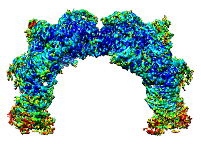 emd_32688.png emd_32688.png | 143.3 KB | ||
| Filedesc metadata | emd-32688.cif.gz | 6.1 KB | ||
| Archive directory |  http://ftp.pdbj.org/pub/emdb/structures/EMD-32688ftp://ftp.pdbj.org/pub/emdb/structures/EMD-32688 http://ftp.pdbj.org/pub/emdb/structures/EMD-32688ftp://ftp.pdbj.org/pub/emdb/structures/EMD-32688 | HTTPS FTP |
-Related structure data
| Related structure data |  7wpqMC  7wppC  7wprC  7wpsC  7wqtC C: citing same article ( M: atomic model generated by this map |
|---|---|
| Similar structure data |


-Links
| EMDB pages | EMDB (EBI/PDBe) / EMDataResource |
|---|---|
| Related items in Molecule of the Month |


-Map
| File | Download / File: emd_32688.map.gz / Format: CCP4 / Size: 47.4 MB / Type: IMAGE STORED AS FLOATING POINT NUMBER (4 BYTES) | ||||||||||||||||||||||||||||||||||||
|---|---|---|---|---|---|---|---|---|---|---|---|---|---|---|---|---|---|---|---|---|---|---|---|---|---|---|---|---|---|---|---|---|---|---|---|---|---|
| Projections & slices | Image control
Images are generated by Spider. generated in cubic-lattice coordinate | ||||||||||||||||||||||||||||||||||||
| Voxel size | X=Y=Z: 0.97 Å | ||||||||||||||||||||||||||||||||||||
| Density |
| ||||||||||||||||||||||||||||||||||||
| Symmetry | Space group: 1 | ||||||||||||||||||||||||||||||||||||
| Details | EMDB XML:
|
 Z (Sec.)
 Y (Row.)
 X (Col.)


-Supplemental data
- Sample components
-Entire : VWF D'D3-D1D2 complex (2 units)
| Entire | Name: VWF D'D3-D1D2 complex (2 units) |
|---|---|
| Components |
|
-Supramolecule #1: VWF D'D3-D1D2 complex (2 units)
| Supramolecule | Name: VWF D'D3-D1D2 complex (2 units) / type: complex / ID: 1 / Parent: 0 / Macromolecule list: #2 |
|---|---|
| Source (natural) | Organism: Homo sapiens (human) |
-Macromolecule #1: von Willebrand antigen 2
| Macromolecule | Name: von Willebrand antigen 2 / type: protein_or_peptide / ID: 1 / Number of copies: 4 / Enantiomer: LEVO |
|---|---|
| Source (natural) | Organism: Homo sapiens (human) |
| Molecular weight | Theoretical: 81.427703 KDa |
| Recombinant expression | Organism: Homo sapiens (human) |
| Sequence | String: AEGTRGRSST ARCSLFGSDF VNTFDGSMYS FAGYCSYLLA GGCQKRSFSI IGDFQNGKRV SLSVYLGEFF DIHLFVNGTV TQGDQRVSM PYASKGLYLE TEAGYYKLSG EAYGFVARID GSGNFQVLLS DRYFNKTCGL CGNFNIFAED DFMTQEGTLT S DPYDFANS ...String: AEGTRGRSST ARCSLFGSDF VNTFDGSMYS FAGYCSYLLA GGCQKRSFSI IGDFQNGKRV SLSVYLGEFF DIHLFVNGTV TQGDQRVSM PYASKGLYLE TEAGYYKLSG EAYGFVARID GSGNFQVLLS DRYFNKTCGL CGNFNIFAED DFMTQEGTLT S DPYDFANS WALSSGEQWC ERASPPSSSC NISSGEMQKG LWEQCQLLKS TSVFARCHPL VDPEPFVALC EKTLCECAGG LE CACPALL EYARTCAQEG MVLYGWTDHS ACSPVCPAGM EYRQCVSPCA RTCQSLHINE MCQERCVDGC SCPEGQLLDE GLC VESTEC PCVHSGKRYP PGTSLSRDCN TCICRNSQWI CSNEECPGEC LVTGQSHFKS FDNRYFTFSG ICQYLLARDC QDHS FSIVI ETVQCADDRD AVCTRSVTVR LPGLHNSLVK LKHGAGVAMD GQDVQLPLLK GDLRIQHTVT ASVRLSYGED LQMDW DGRG RLLVKLSPVY AGKTCGLCGN YNGNQGDDFL TPSGLAEPRV EDFGNAWKLH GDCQDLQKQH SDPCALNPRM TRFSEE ACA VLTSPTFEAC HRAVSPLPYL RNCRYDVCSC SDGRECLCGA LASYAAACAG RGVRVAWREP GRCELNCPKG QVYLQCG TP CNLTCRSLSY PDEECNEACL EGCFCPPGLY MDERGDCVPK AQCPCYYDGE IFQPEDIFSD HHTMCYCEDG FMHCTMSG V PGSLLPDAVL SSPLSHRSKR UniProtKB: von Willebrand factor |
-Macromolecule #2: von Willebrand factor
| Macromolecule | Name: von Willebrand factor / type: protein_or_peptide / ID: 2 / Number of copies: 4 / Enantiomer: LEVO |
|---|---|
| Source (natural) | Organism: Homo sapiens (human) |
| Molecular weight | Theoretical: 54.232809 KDa |
| Recombinant expression | Organism: Homo sapiens (human) |
| Sequence | String: SLSCRPPMVK LVCPADNLRA EGLECTKTCQ NYDLECMSMG CVSGCLCPPG MVRHENRCVA LERCPCFHQG KEYAPGETVK IGCNTCVCQ DRKWNCTDHV CDATCSTIGM AHYLTFDGLK YLFPGECQYV LVQDYCGSNP GTFRILVGNK GCSHPSVKCK K RVTILVEG ...String: SLSCRPPMVK LVCPADNLRA EGLECTKTCQ NYDLECMSMG CVSGCLCPPG MVRHENRCVA LERCPCFHQG KEYAPGETVK IGCNTCVCQ DRKWNCTDHV CDATCSTIGM AHYLTFDGLK YLFPGECQYV LVQDYCGSNP GTFRILVGNK GCSHPSVKCK K RVTILVEG GEIELFDGEV NVKRPMKDET HFEVVESGRY IILLLGKALS VVWDRHLSIS VVLKQTYQEK VCGLCGNFDG IQ NNDLTSS NLQVEEDPVD FGNSWKVSSQ CADTRKVPLD SSPATCHNNI MKQTMVDSSC RILTSDVFQD CNKLVDPEPY LDV CIYDTC SCESIGDCAC FCDTIAAYAH VCAQHGKVVT WRTATLCPQS CEERNLRENG YECEWRYNSC APACQVTCQH PEPL ACPVQ CVEGCHAHCP PGKILDELLQ TCVDPEDCPV CEVAGRRFAS GKKVTLNPSD PEHCQICHCD VVNLTCEACQ EPGGL VVPP HHHHHH UniProtKB: von Willebrand factor |
-Macromolecule #3: 2-acetamido-2-deoxy-beta-D-glucopyranose
| Macromolecule | Name: 2-acetamido-2-deoxy-beta-D-glucopyranose / type: ligand / ID: 3 / Number of copies: 20 / Formula: NAG |
|---|---|
| Molecular weight | Theoretical: 221.208 Da |
| Chemical component information |  ChemComp-NAG: |
-Macromolecule #4: CALCIUM ION
| Macromolecule | Name: CALCIUM ION / type: ligand / ID: 4 / Number of copies: 16 / Formula: CA |
|---|---|
| Molecular weight | Theoretical: 40.078 Da |
-Experimental details
-Structure determination
| Method | cryo EM |
|---|---|
 Processing Processing | single particle reconstruction |
| Aggregation state | particle |
-Sample preparation
| Buffer | pH: 7.4 |
|---|---|
| Vitrification | Cryogen name: ETHANE |
- Electron microscopy
| Microscope | FEI TITAN KRIOS |
|---|---|
| Image recording | Film or detector model: GATAN K3 BIOQUANTUM (6k x 4k) / Average electron dose: 50.0 e/Å2 |
| Electron beam | Acceleration voltage: 300 kV / Electron source:  FIELD EMISSION GUN FIELD EMISSION GUN |
| Electron optics | Illumination mode: OTHER / Imaging mode: OTHER / Nominal defocus max: 2.0 µm / Nominal defocus min: 1.0 µm |
| Experimental equipment |  Model: Titan Krios / Image courtesy: FEI Company |
-Image processing
| Startup model | Type of model: INSILICO MODEL |
|---|---|
| Final reconstruction | Applied symmetry - Point group: C1 (asymmetric) / Resolution.type: BY AUTHOR / Resolution: 3.267 Å / Resolution method: FSC 0.143 CUT-OFF / Number images used: 539243 |
| Initial angle assignment | Type: MAXIMUM LIKELIHOOD |
| Final angle assignment | Type: MAXIMUM LIKELIHOOD |
