- EMDB-32067: Cryo-EM structure of the human ATP13A2 (E1P-ADP state) -
+
Open data
ID or keywords:
Loading...
-
Basic information
Entry
Database: EMDB / ID: EMD-32067
Title
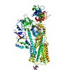
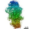
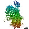
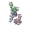
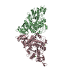
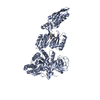
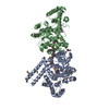
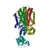
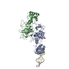
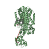
Cryo-EM structure of the human ATP13A2 (E1P-ADP state)
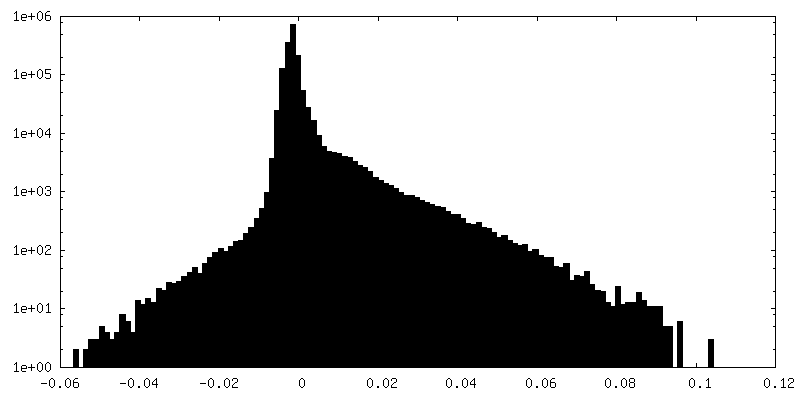
Map data
postprocess_masked.mrc from Relion
Sample
Complex: ATP13A2
Protein or peptide: Polyamine-transporting ATPase 13A2
Ligand: MAGNESIUM ION
Ligand: ADENOSINE-5'-DIPHOSPHATE
Ligand: 2-acetamido-2-deoxy-beta-D-glucopyranose
Ligand: TETRAFLUOROALUMINATE ION
Keywords
ATP13A2 / PARK9 / P-type ATPase / MEMBRANE PROTEIN
Function / homology
Function and homology information
polyamine transmembrane transport / spermine transmembrane transport / polyamine transmembrane transporter activity / ABC-type polyamine transporter activity / regulation of autophagosome size / extracellular exosome biogenesis / negative regulation of lysosomal protein catabolic process / regulation of chaperone-mediated autophagy / P-type ion transporter activity / regulation of lysosomal protein catabolic process ...polyamine transmembrane transport / spermine transmembrane transport / polyamine transmembrane transporter activity / ABC-type polyamine transporter activity / regulation of autophagosome size / extracellular exosome biogenesis / negative regulation of lysosomal protein catabolic process / regulation of chaperone-mediated autophagy / P-type ion transporter activity / regulation of lysosomal protein catabolic process / regulation of autophagy of mitochondrion / intracellular monoatomic cation homeostasis / autophagosome-lysosome fusion / autophagosome organization / protein localization to lysosome / positive regulation of exosomal secretion / ATPase-coupled monoatomic cation transmembrane transporter activity / multivesicular body membrane / phosphatidic acid binding / intracellular zinc ion homeostasis / cupric ion binding / regulation of mitochondrion organization / regulation of protein localization to nucleus / phosphatidylinositol-3,5-bisphosphate binding / Translocases; Catalysing the translocation of other compounds; Linked to the hydrolysis of a nucleoside triphosphate / lysosomal transport / regulation of intracellular protein transport / lipid homeostasis / cellular response to zinc ion / autophagosome membrane / Ion transport by P-type ATPases / regulation of macroautophagy / transport vesicle / cellular response to manganese ion / multivesicular body / lysosomal lumen / autophagosome / regulation of neuron apoptotic process / positive regulation of protein secretion / autophagy / intracellular calcium ion homeostasis / late endosome / late endosome membrane / manganese ion binding / cellular response to oxidative stress / monoatomic ion transmembrane transport / vesicle / intracellular iron ion homeostasis / lysosome / neuron projection / lysosomal membrane / neuronal cell body / positive regulation of gene expression / ATP hydrolysis activity / zinc ion binding / ATP binding / membrane Similarity search - Function
Japan Agency for Medical Research and Development (AMED)
JP19am01011115
Japan
Citation
Journal: Mol Cell / Year: 2021 Title: Cryo-EM reveals mechanistic insights into lipid-facilitated polyamine export by human ATP13A2. Authors: Atsuhiro Tomita / Takashi Daiho / Tsukasa Kusakizako / Keitaro Yamashita / Satoshi Ogasawara / Takeshi Murata / Tomohiro Nishizawa / Osamu Nureki / Abstract: The cytoplasmic polyamine maintains cellular homeostasis by chelating toxic metal cations, regulating transcriptional activity, and protecting DNA. ATP13A2 was identified as a lysosomal polyamine ...The cytoplasmic polyamine maintains cellular homeostasis by chelating toxic metal cations, regulating transcriptional activity, and protecting DNA. ATP13A2 was identified as a lysosomal polyamine exporter responsible for polyamine release into the cytosol, and its dysfunction is associated with Alzheimer's disease and other neural degradation diseases. ATP13A2 belongs to the P5 subfamily of the P-type ATPase family, but its mechanisms remain unknown. Here, we report the cryoelectron microscopy (cryo-EM) structures of human ATP13A2 under four different conditions, revealing the structural coupling between the polyamine binding and the dephosphorylation. Polyamine is bound at the luminal tunnel and recognized through numerous electrostatic and π-cation interactions, explaining its broad specificity. The unique N-terminal domain is anchored to the lipid membrane to stabilize the E2P conformation, thereby accelerating the E1P-to-E2P transition. These findings reveal the distinct mechanism of P5B ATPases, thereby paving the way for neuroprotective therapy by activating ATP13A2.
History
Deposition
Oct 17, 2021
-
Header (metadata) release
Dec 29, 2021
-
Map release
Dec 29, 2021
-
Update
Oct 30, 2024
-
Current status
Oct 30, 2024
Processing site: PDBj / Status: Released
-
Structure visualization
Movie
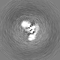
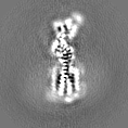
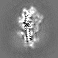
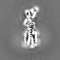
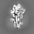
Surface view with section colored by density value
EMPIAR-10975 (Title: Cryo-EM structure of the human ATP13A2 / Data size: 3.8 TB Data #1: Unaligned movies for E1-ATP state [micrographs - multiframe] Data #2: Unaligned movies for E1P-ADP state [micrographs - multiframe] Data #3: Unaligned movies for SPM-bound E2P state [micrographs - multiframe] Data #4: Unaligned movies for SPM-bound E2Pi state [micrographs - multiframe])
Name: Polyamine-transporting ATPase 13A2 / type: protein_or_peptide / ID: 1 / Number of copies: 1 / Enantiomer: LEVO EC number: Translocases; Catalysing the translocation of other compounds; Linked to the hydrolysis of a nucleoside triphosphate
In the structure databanks used in Yorodumi, some data are registered as the other names, "COVID-19 virus" and "2019-nCoV". Here are the details of the virus and the list of structure data.
Jan 31, 2019. EMDB accession codes are about to change! (news from PDBe EMDB page)
EMDB accession codes are about to change! (news from PDBe EMDB page)
The allocation of 4 digits for EMDB accession codes will soon come to an end. Whilst these codes will remain in use, new EMDB accession codes will include an additional digit and will expand incrementally as the available range of codes is exhausted. The current 4-digit format prefixed with “EMD-” (i.e. EMD-XXXX) will advance to a 5-digit format (i.e. EMD-XXXXX), and so on. It is currently estimated that the 4-digit codes will be depleted around Spring 2019, at which point the 5-digit format will come into force.
The EM Navigator/Yorodumi systems omit the EMD- prefix.
Related info.:Q: What is EMD? / ID/Accession-code notation in Yorodumi/EM Navigator
Yorodumi is a browser for structure data from EMDB, PDB, SASBDB, etc.
This page is also the successor to EM Navigator detail page, and also detail information page/front-end page for Omokage search.
The word "yorodu" (or yorozu) is an old Japanese word meaning "ten thousand". "mi" (miru) is to see.
Related info.:EMDB / PDB / SASBDB / Comparison of 3 databanks / Yorodumi Search / Aug 31, 2016. New EM Navigator & Yorodumi / Yorodumi Papers / Jmol/JSmol / Function and homology information / Changes in new EM Navigator and Yorodumi
 Movie
Movie Controller


 Open data
 Basic information
 Map data
 Sample
 Keywords
 Function and homology information
 Homo sapiens (human)
 Authors
 Japan, 3 items
  Citation
 Structure visualization
 Downloads & links
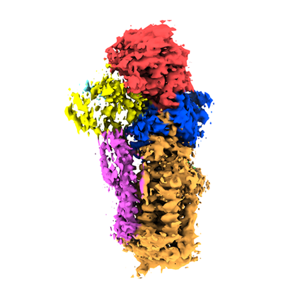 emd_32067.png
 http://ftp.pdbj.org/pub/emdb/structures/EMD-32067
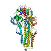


 Z (Sec.)
 Y (Row.)
 X (Col.)


 Sample components


 Processing
 Electron microscopy
 FIELD EMISSION GUN
