 Movie
Movie Controller

[English] 日本語
 Yorodumi
- PDB-5ykb: The N253F mutant structure of trehalose synthase from Deinococcus... -
+ Open data

- Basic information
| Entry | Database: PDB / ID: 5ykb | |||||||||
|---|---|---|---|---|---|---|---|---|---|---|
| Title | The N253F mutant structure of trehalose synthase from Deinococcus radiodurans reveals an open active-site conformation | |||||||||
 Components Components | Trehalose synthase | |||||||||
 Keywords Keywords | ISOMERASE / trehalose synthase / glycoside hydrolase family 13 | |||||||||
| Function / homology |  Function and homology information Function and homology informationmaltose alpha-D-glucosyltransferase / maltose alpha-D-glucosyltransferase activity / carbohydrate metabolic process / metal ion binding Similarity search - Function | |||||||||
| Biological species |  Deinococcus radiodurans str. R1 (radioresistant) Deinococcus radiodurans str. R1 (radioresistant) | |||||||||
| Method |  X-RAY DIFFRACTION / SYNCHROTRON / MOLECULAR REPLACEMENT / Resolution: 2.76 Å X-RAY DIFFRACTION / SYNCHROTRON / MOLECULAR REPLACEMENT / Resolution: 2.76 Å | |||||||||
 Authors Authors | Chow, S.Y. / Hsieh, Y.C. / Liaw, S.H. | |||||||||
| Funding support |  Taiwan, 1items Taiwan, 1items
| |||||||||
 Citation Citation | Journal: Acta Crystallogr F Struct Biol Commun / Year: 2017 Title: The N253F mutant structure of trehalose synthase from Deinococcus radiodurans reveals an open active-site topology Authors: Chow, S.Y. / Wang, Y.L. / Hsieh, Y.C. / Lee, G.C. / Liaw, S.H. | |||||||||
| History |
|
- Structure visualization
| Structure viewer | Molecule: MolmilJmol/JSmol |
|---|
- Downloads & links
-Download
| PDBx/mmCIF format | 5ykb.cif.gz | 427.5 KB | Display | PDBx/mmCIF format |
|---|---|---|---|---|
| PDB format | pdb5ykb.ent.gz | 347.5 KB | Display | PDB format |
| PDBx/mmJSON format | 5ykb.json.gz | Tree view | PDBx/mmJSON format | |
| Others |  Other downloads Other downloads |
-Validation report
| Arichive directory | https://data.pdbj.org/pub/pdb/validation_reports/yk/5ykbftp://data.pdbj.org/pub/pdb/validation_reports/yk/5ykb | HTTPS FTP |
|---|
-Related structure data
| Related structure data |  4tvuS S: Starting model for refinement |
|---|---|
| Similar structure data |


-Links
 PDBj
PDBj

- Assembly
| Deposited unit | 
| ||||||||||||||||||||||||||||||||||||||||||||||||||||||||||||||||||||||||||||||||||||||||||||||||||
|---|---|---|---|---|---|---|---|---|---|---|---|---|---|---|---|---|---|---|---|---|---|---|---|---|---|---|---|---|---|---|---|---|---|---|---|---|---|---|---|---|---|---|---|---|---|---|---|---|---|---|---|---|---|---|---|---|---|---|---|---|---|---|---|---|---|---|---|---|---|---|---|---|---|---|---|---|---|---|---|---|---|---|---|---|---|---|---|---|---|---|---|---|---|---|---|---|---|---|---|
| 1 | 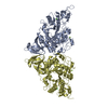
| ||||||||||||||||||||||||||||||||||||||||||||||||||||||||||||||||||||||||||||||||||||||||||||||||||
| 2 | 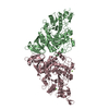
| ||||||||||||||||||||||||||||||||||||||||||||||||||||||||||||||||||||||||||||||||||||||||||||||||||
| Unit cell |
| ||||||||||||||||||||||||||||||||||||||||||||||||||||||||||||||||||||||||||||||||||||||||||||||||||
| Noncrystallographic symmetry (NCS) | NCS domain:
NCS domain segments: Component-ID: _ / Beg auth comp-ID: PRO / Beg label comp-ID: PRO / End auth comp-ID: ASN / End label comp-ID: ASN / Refine code: _ / Auth seq-ID: 6 - 552 / Label seq-ID: 8 - 554
NCS ensembles :
|
-Components
| #1: Protein | Mass: 65020.914 Da / Num. of mol.: 4 / Mutation: R97W,N253F,T313I,I380V Source method: isolated from a genetically manipulated source Source: (gene. exp.) Deinococcus radiodurans str. R1 (radioresistant)Strain: R1 / Production host: References: UniProt: I3NX86, maltose alpha-D-glucosyltransferase #2: Chemical | ChemComp-CA /   Mass: 40.078 Da / Num. of mol.: 4 / Source method: obtained synthetically / Formula: Ca Mass: 40.078 Da / Num. of mol.: 4 / Source method: obtained synthetically / Formula: Ca#3: Chemical | ChemComp-MG /   Mass: 24.305 Da / Num. of mol.: 4 / Source method: obtained synthetically / Formula: Mg Mass: 24.305 Da / Num. of mol.: 4 / Source method: obtained synthetically / Formula: Mg#4: Water | ChemComp-HOH / |  Mass: 18.015 Da / Num. of mol.: 237 / Source method: isolated from a natural source / Formula: H2O Mass: 18.015 Da / Num. of mol.: 237 / Source method: isolated from a natural source / Formula: H2O |
|---|
-Experimental details
-Experiment
| Experiment | Method: X-RAY DIFFRACTION / Number of used crystals: 1 |
|---|
- Sample preparation
| Crystal | Density Matthews: 2.55 Å3/Da / Density % sol: 51.78 % |
|---|---|
| Crystal grow | Temperature: 288 K / Method: vapor diffusion, hanging drop / pH: 7 Details: 7% PEG 4000, 0.2M sodium acetate trihydrate, 0.3M Tris-HCl (pH 7.0) |
-Data collection
| Diffraction | Mean temperature: 110 K |
|---|---|
| Diffraction source | Source: SYNCHROTRON / Site: NSRRC / Beamline: BL15A1 / Wavelength: 1 Å |
| Detector | Type: RAYONIX MX300HE / Detector: CCD / Date: May 10, 2014 |
| Radiation | Protocol: SINGLE WAVELENGTH / Monochromatic (M) / Laue (L): M / Scattering type: x-ray |
| Radiation wavelength | Wavelength: 1 Å / Relative weight: 1 |
| Reflection | Resolution: 2.76→30 Å / Num. obs: 67645 / % possible obs: 99.9 % / Redundancy: 4.5 % / CC1/2: 0.993 / Net I/σ(I): 12.3 |
| Reflection shell | Resolution: 2.76→2.86 Å / Redundancy: 4.4 % / Mean I/σ(I) obs: 1.8 / Num. unique obs: 6642 / CC1/2: 0.606 / % possible all: 99.3 |
- Processing
| Software |
| |||||||||||||||||||||||||||||||||||||||||||||||||||||||||||||||||
|---|---|---|---|---|---|---|---|---|---|---|---|---|---|---|---|---|---|---|---|---|---|---|---|---|---|---|---|---|---|---|---|---|---|---|---|---|---|---|---|---|---|---|---|---|---|---|---|---|---|---|---|---|---|---|---|---|---|---|---|---|---|---|---|---|---|---|
| Refinement | Method to determine structure: MOLECULAR REPLACEMENT Starting model: 4TVU Resolution: 2.76→30 Å / Cor.coef. Fo:Fc: 0.939 / Cor.coef. Fo:Fc free: 0.884 / SU B: 14.965 / SU ML: 0.298 / Cross valid method: THROUGHOUT / σ(F): 0 / ESU R Free: 0.374 Details: HYDROGENS HAVE BEEN ADDED IN THE RIDING POSITIONS U VALUES : REFINED INDIVIDUALLY
| |||||||||||||||||||||||||||||||||||||||||||||||||||||||||||||||||
| Solvent computation | Ion probe radii: 0.8 Å / Shrinkage radii: 0.8 Å / VDW probe radii: 1.2 Å | |||||||||||||||||||||||||||||||||||||||||||||||||||||||||||||||||
| Displacement parameters | Biso max: 136.58 Å2 / Biso mean: 50.759 Å2 / Biso min: 2 Å2
| |||||||||||||||||||||||||||||||||||||||||||||||||||||||||||||||||
| Refinement step | Cycle: final / Resolution: 2.76→30 Å
| |||||||||||||||||||||||||||||||||||||||||||||||||||||||||||||||||
| Refine LS restraints |
| |||||||||||||||||||||||||||||||||||||||||||||||||||||||||||||||||
| Refine LS restraints NCS | Refine-ID: X-RAY DIFFRACTION / Type: interatomic distance / Weight position: 0.05
| |||||||||||||||||||||||||||||||||||||||||||||||||||||||||||||||||
| LS refinement shell | Resolution: 2.761→2.832 Å / Rfactor Rfree error: 0 / Total num. of bins used: 20
|
