- EMDB-32033: 14pf microtubule decorated with EML1-GFP -
+
Open data
ID or keywords:
Loading...
-
Basic information
Entry
Database: EMDB / ID: EMD-32033
Title
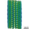
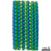
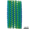
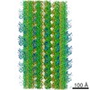
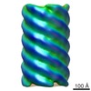
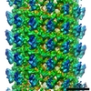
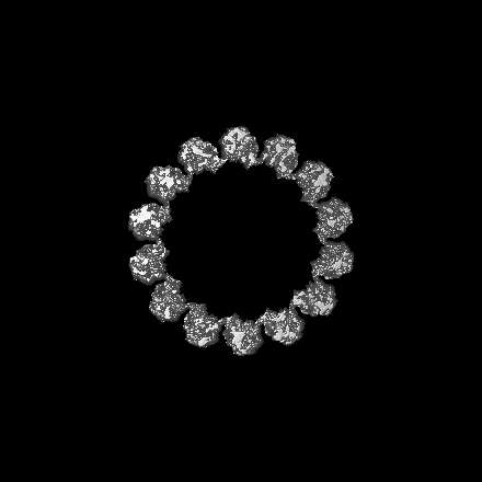
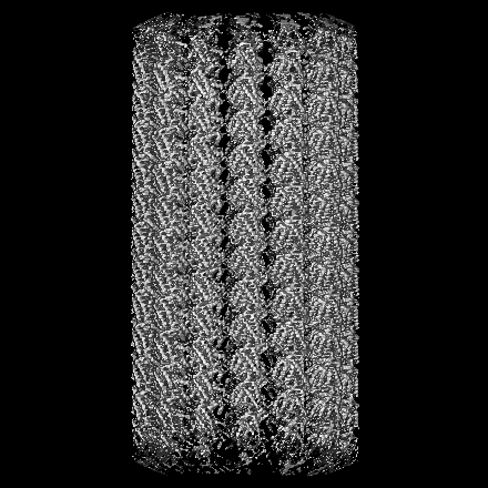
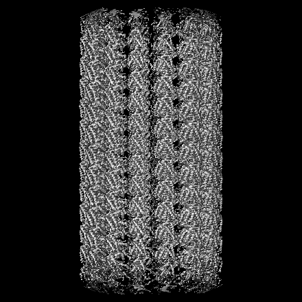
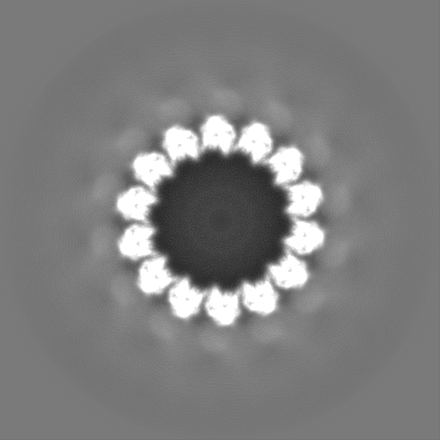
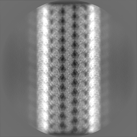
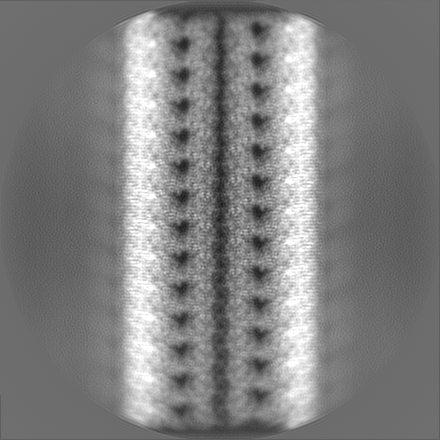
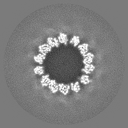
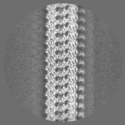
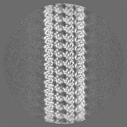
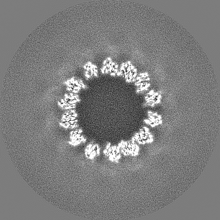
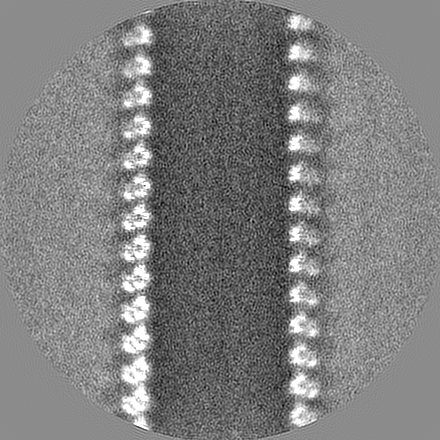
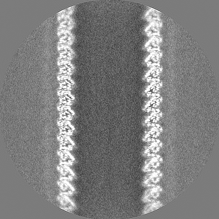
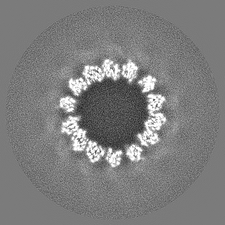
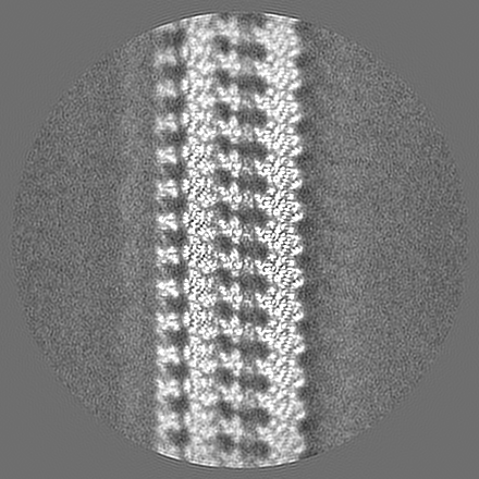
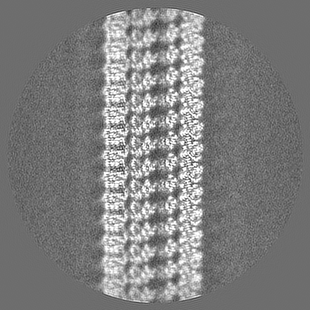
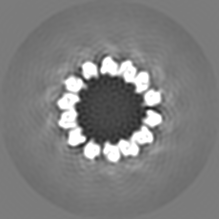
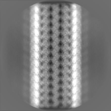
14pf microtubule decorated with EML1-GFP
Map data
Refined map of 14pf microtubule bound to EML1-GFP
Sample
Complex: Complex of microtubule with HEK293 cell lysate containing over expressed EML1-GFP
Protein or peptide: Tubulin alpha-1B chain
Protein or peptide: Tubulin beta chain
Keywords
Cytoskeleton / Echinoderm microtubule-associated protein-like 1 (EML1) / Microtubule associated protein (MAP) / STRUCTURAL PROTEIN
Function / homology
Function and homology information
odontoblast differentiation / Cilium Assembly / Post-chaperonin tubulin folding pathway / cytoskeleton-dependent intracellular transport / Carboxyterminal post-translational modifications of tubulin / Microtubule-dependent trafficking of connexons from Golgi to the plasma membrane / Sealing of the nuclear envelope (NE) by ESCRT-III / Intraflagellar transport / Formation of tubulin folding intermediates by CCT/TriC / Gap junction assembly ...odontoblast differentiation / Cilium Assembly / Post-chaperonin tubulin folding pathway / cytoskeleton-dependent intracellular transport / Carboxyterminal post-translational modifications of tubulin / Microtubule-dependent trafficking of connexons from Golgi to the plasma membrane / Sealing of the nuclear envelope (NE) by ESCRT-III / Intraflagellar transport / Formation of tubulin folding intermediates by CCT/TriC / Gap junction assembly / Kinesins / Assembly and cell surface presentation of NMDA receptors / COPI-independent Golgi-to-ER retrograde traffic / GTPase activating protein binding / natural killer cell mediated cytotoxicity / COPI-dependent Golgi-to-ER retrograde traffic / nuclear envelope lumen / regulation of synapse organization / Recycling pathway of L1 / RHOH GTPase cycle / MHC class I protein binding / microtubule-based process / RHO GTPases activate IQGAPs / Hedgehog 'off' state / intercellular bridge / COPI-mediated anterograde transport / cytoplasmic microtubule / Activation of AMPK downstream of NMDARs / cellular response to interleukin-4 / ciliary tip / Loss of Nlp from mitotic centrosomes / Loss of proteins required for interphase microtubule organization from the centrosome / Recruitment of mitotic centrosome proteins and complexes / MHC class II antigen presentation / Recruitment of NuMA to mitotic centrosomes / Anchoring of the basal body to the plasma membrane / HSP90 chaperone cycle for steroid hormone receptors (SHR) in the presence of ligand / Mitotic Prometaphase / EML4 and NUDC in mitotic spindle formation / AURKA Activation by TPX2 / Resolution of Sister Chromatid Cohesion / Translocation of SLC2A4 (GLUT4) to the plasma membrane / sperm end piece / sperm principal piece / RHO GTPases Activate Formins / PKR-mediated signaling / microtubule cytoskeleton organization / structural constituent of cytoskeleton / HCMV Early Events / cytoplasmic ribonucleoprotein granule / Aggrephagy / mitotic spindle / azurophil granule lumen / The role of GTSE1 in G2/M progression after G2 checkpoint / Separation of Sister Chromatids / Regulation of PLK1 Activity at G2/M Transition / mitotic cell cycle / double-stranded RNA binding / microtubule cytoskeleton / cell body / Potential therapeutics for SARS / microtubule / Hydrolases; Acting on acid anhydrides; Acting on GTP to facilitate cellular and subcellular movement / cytoskeleton / cilium / membrane raft / protein domain specific binding / cell division / GTPase activity / ubiquitin protein ligase binding / Neutrophil degranulation / GTP binding / protein-containing complex binding / structural molecule activity / protein-containing complex / extracellular exosome / extracellular region / metal ion binding / nucleus / cytosol / cytoplasm Similarity search - Function
Journal: Nat Cell Biol / Year: 2022 Title: Lysate-based pipeline to characterize microtubule-associated proteins uncovers unique microtubule behaviours. Authors: A S Jijumon / Satish Bodakuntla / Mariya Genova / Mamata Bangera / Violet Sackett / Laetitia Besse / Fatlinda Maksut / Veronique Henriot / Maria M Magiera / Minhajuddin Sirajuddin / Carsten Janke / Abstract: The microtubule cytoskeleton forms complex macromolecular assemblies with a range of microtubule-associated proteins (MAPs) that have fundamental roles in cell architecture, division and motility. ...The microtubule cytoskeleton forms complex macromolecular assemblies with a range of microtubule-associated proteins (MAPs) that have fundamental roles in cell architecture, division and motility. Determining how an individual MAP modulates microtubule behaviour is an important step in understanding the physiological roles of various microtubule assemblies. To characterize how MAPs control microtubule properties and functions, we developed an approach allowing for medium-throughput analyses of MAPs in cell-free conditions using lysates of mammalian cells. Our pipeline allows for quantitative as well as ultrastructural analyses of microtubule-MAP assemblies. Analysing 45 bona fide and potential mammalian MAPs, we uncovered previously unknown activities that lead to distinct and unique microtubule behaviours such as microtubule coiling or hook formation, or liquid-liquid phase separation along the microtubule lattice that initiates microtubule branching. We have thus established a powerful tool for a thorough characterization of a wide range of MAPs and MAP variants, thus opening avenues for the determination of mechanisms underlying their physiological roles and pathological implications.
History
Deposition
Oct 7, 2021
-
Header (metadata) release
Dec 1, 2021
-
Map release
Dec 1, 2021
-
Update
Dec 13, 2023
-
Current status
Dec 13, 2023
Processing site: PDBj / Status: Released
-
Structure visualization
Movie
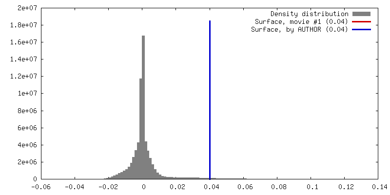
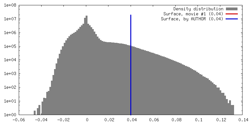
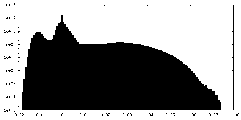
Surface view with section colored by density value
Entire : Complex of microtubule with HEK293 cell lysate containing over ex...
Entire
Name: Complex of microtubule with HEK293 cell lysate containing over expressed EML1-GFP
Components
Complex: Complex of microtubule with HEK293 cell lysate containing over expressed EML1-GFP
Protein or peptide: Tubulin alpha-1B chain
Protein or peptide: Tubulin beta chain
-
Supramolecule #1: Complex of microtubule with HEK293 cell lysate containing over ex...
Supramolecule
Name: Complex of microtubule with HEK293 cell lysate containing over expressed EML1-GFP type: complex / ID: 1 / Parent: 0 / Macromolecule list: all
Source (natural)
Organism: Homo sapiens (human)
-
Macromolecule #1: Tubulin alpha-1B chain
Macromolecule
Name: Tubulin alpha-1B chain / type: protein_or_peptide / ID: 1 Details: The reconstruction contains a mix of tubulin from goat brain (nucleating seeds) and tubulin from HEK293 cellular lysate so will have a mixture of both sequences. Enantiomer: LEVO
Name: Tubulin beta chain / type: protein_or_peptide / ID: 2 Details: The reconstruction contains a mix of tubulin from goat brain (nucleating seeds) and tubulin from HEK293 cellular lysate so will have a mixture of both sequences. Sequence of goat brain beta ...Details: The reconstruction contains a mix of tubulin from goat brain (nucleating seeds) and tubulin from HEK293 cellular lysate so will have a mixture of both sequences. Sequence of goat brain beta tubulin can be accessed using Uniprot ID A0A452G3J7 Enantiomer: LEVO
Model: Quantifoil R1.2/1.3 / Material: GOLD / Mesh: 300 / Support film - Material: CARBON / Support film - topology: HOLEY / Pretreatment - Type: GLOW DISCHARGE / Pretreatment - Time: 90 sec. / Pretreatment - Atmosphere: AIR / Pretreatment - Pressure: 0.025 kPa
Vitrification
Cryogen name: ETHANE / Chamber humidity: 100 % / Chamber temperature: 298 K / Instrument: FEI VITROBOT MARK IV Details: GMPCPP MT seeds in warm BRB80 applied to the grid followed by second application of pre-warmed lysate containing over expressed EML1-GFP. After an incubation time of 20 seconds, grid blotted ...Details: GMPCPP MT seeds in warm BRB80 applied to the grid followed by second application of pre-warmed lysate containing over expressed EML1-GFP. After an incubation time of 20 seconds, grid blotted for 3 seconds before plunging into liquid ethane..
Details
Goat brain tubulin was polymerized with 1mM GMPCPP at 310K for 2 hours, spun on warm 50 percent BRB80 sucrose cushion and resuspended in twice the volume of warm BRB80 buffer
-
Electron microscopy
Microscope
FEI TITAN KRIOS
Image recording
Film or detector model: FEI FALCON III (4k x 4k) / Detector mode: INTEGRATING / Digitization - Dimensions - Width: 4096 pixel / Digitization - Dimensions - Height: 4096 pixel / Number grids imaged: 1 / Number real images: 1143 / Average exposure time: 1.5 sec. / Average electron dose: 44.17 e/Å2 / Details: 30 frames
Electron beam
Acceleration voltage: 300 kV / Electron source: FIELD EMISSION GUN
Electron optics
C2 aperture diameter: 50.0 µm / Calibrated magnification: 101449 / Illumination mode: OTHER / Imaging mode: OTHER / Cs: 2.7 mm / Nominal magnification: 59000
In the structure databanks used in Yorodumi, some data are registered as the other names, "COVID-19 virus" and "2019-nCoV". Here are the details of the virus and the list of structure data.
Jan 31, 2019. EMDB accession codes are about to change! (news from PDBe EMDB page)
EMDB accession codes are about to change! (news from PDBe EMDB page)
The allocation of 4 digits for EMDB accession codes will soon come to an end. Whilst these codes will remain in use, new EMDB accession codes will include an additional digit and will expand incrementally as the available range of codes is exhausted. The current 4-digit format prefixed with “EMD-” (i.e. EMD-XXXX) will advance to a 5-digit format (i.e. EMD-XXXXX), and so on. It is currently estimated that the 4-digit codes will be depleted around Spring 2019, at which point the 5-digit format will come into force.
The EM Navigator/Yorodumi systems omit the EMD- prefix.
Related info.:Q: What is EMD? / ID/Accession-code notation in Yorodumi/EM Navigator
Yorodumi is a browser for structure data from EMDB, PDB, SASBDB, etc.
This page is also the successor to EM Navigator detail page, and also detail information page/front-end page for Omokage search.
The word "yorodu" (or yorozu) is an old Japanese word meaning "ten thousand". "mi" (miru) is to see.
Related info.:EMDB / PDB / SASBDB / Comparison of 3 databanks / Yorodumi Search / Aug 31, 2016. New EM Navigator & Yorodumi / Yorodumi Papers / Jmol/JSmol / Function and homology information / Changes in new EM Navigator and Yorodumi
 Movie
Movie Controller


 Open data
 Basic information
 Map data
 Sample
 Keywords
 Function and homology information
 Homo sapiens (human)
 Authors
 Citation


 Structure visualization
 Downloads & links
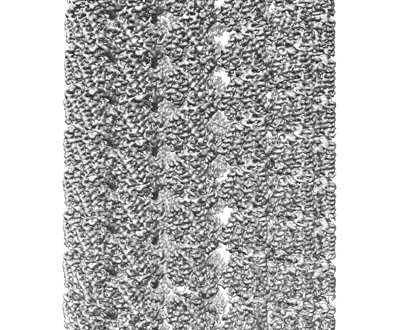 emd_32033.png
 http://ftp.pdbj.org/pub/emdb/structures/EMD-32033
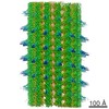


 Z (Sec.)
 Y (Row.)
 X (Col.)


 Sample components
 Processing
 Electron microscopy
 FIELD EMISSION GUN

