 Movie
Movie Controller

[English] 日本語
 Yorodumi
- EMDB-1974: Electron Cryotomography of Measles Virus Reveals how Matrix Prote... -
+ Open data

- Basic information
| Entry | Database: EMDB / ID: EMD-1974 | |||||||||
|---|---|---|---|---|---|---|---|---|---|---|
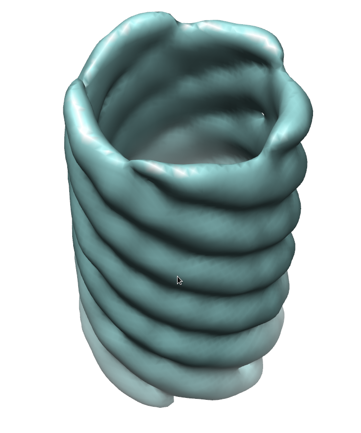
| Title | Electron Cryotomography of Measles Virus Reveals how Matrix Protein Coats the Ribonucleocapsid Within Intact Virions. | |||||||||
 Map data Map data | Measles virus matrix filament | |||||||||
 Sample Sample |
| |||||||||
 Keywords Keywords | measles / matrix / nucleocapsid / ribonucleocapsid / RNP / MCNC | |||||||||
| Biological species |   Measles virus Measles virus | |||||||||
| Method | subtomogram averaging / cryo EM / Resolution: 44.0 Å | |||||||||
 Authors Authors | Liljeroos L / Huiskonen JT / Ora A / Susi P / Butcher SJ | |||||||||
 Citation Citation | Journal: Proc Natl Acad Sci U S A / Year: 2011 Title: Electron cryotomography of measles virus reveals how matrix protein coats the ribonucleocapsid within intact virions. Authors: Lassi Liljeroos / Juha T Huiskonen / Ari Ora / Petri Susi / Sarah J Butcher /  Abstract: Measles virus is a highly infectious, enveloped, pleomorphic virus. We combined electron cryotomography with subvolume averaging and immunosorbent electron microscopy to characterize the 3D ...Measles virus is a highly infectious, enveloped, pleomorphic virus. We combined electron cryotomography with subvolume averaging and immunosorbent electron microscopy to characterize the 3D ultrastructure of the virion. We show that the matrix protein forms helices coating the helical ribonucleocapsid rather than coating the inner leaflet of the membrane, as previously thought. The ribonucleocapsid is folded into tight bundles through matrix-matrix interactions. The implications for virus assembly are that the matrix already tightly interacts with the ribonucleocapsid in the cytoplasm, providing a structural basis for the previously observed regulation of RNA transcription by the matrix protein. Next, the matrix-covered ribonucleocapsids are transported to the plasma membrane, where the matrix interacts with the envelope glycoproteins during budding. These results are relevant to the nucleocapsid organization and budding of other paramyxoviruses, where isolated matrix has been observed to form helices. | |||||||||
| History |
|
- Structure visualization
| Movie |
 Movie viewer Movie viewer |
|---|---|
| Structure viewer | EM map: SurfViewMolmilJmol/JSmol |
| Supplemental images |
- Downloads & links
-EMDB archive
| Map data | emd_1974.map.gz | 1.4 MB | EMDB map data format | |
|---|---|---|---|---|
| Header (meta data) | emd-1974-v30.xmlemd-1974.xml | 8.2 KB 8.2 KB | Display Display | EMDB header |
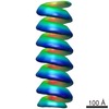
| Images |  emd1974.png emd1974.png | 237.4 KB | ||
| Archive directory |  http://ftp.pdbj.org/pub/emdb/structures/EMD-1974ftp://ftp.pdbj.org/pub/emdb/structures/EMD-1974 http://ftp.pdbj.org/pub/emdb/structures/EMD-1974ftp://ftp.pdbj.org/pub/emdb/structures/EMD-1974 | HTTPS FTP |
-Related structure data
-Links
| EMDB pages | EMDB (EBI/PDBe) / EMDataResource |
|---|
-Map
| File | Download / File: emd_1974.map.gz / Format: CCP4 / Size: 1.4 MB / Type: IMAGE STORED AS FLOATING POINT NUMBER (4 BYTES) | ||||||||||||||||||||||||||||||||||||||||||||||||||||||||||||||||||||
|---|---|---|---|---|---|---|---|---|---|---|---|---|---|---|---|---|---|---|---|---|---|---|---|---|---|---|---|---|---|---|---|---|---|---|---|---|---|---|---|---|---|---|---|---|---|---|---|---|---|---|---|---|---|---|---|---|---|---|---|---|---|---|---|---|---|---|---|---|---|
| Annotation | Measles virus matrix filament | ||||||||||||||||||||||||||||||||||||||||||||||||||||||||||||||||||||
| Projections & slices | Image control
Images are generated by Spider. | ||||||||||||||||||||||||||||||||||||||||||||||||||||||||||||||||||||
| Voxel size | X=Y=Z: 7.68 Å | ||||||||||||||||||||||||||||||||||||||||||||||||||||||||||||||||||||
| Density |
| ||||||||||||||||||||||||||||||||||||||||||||||||||||||||||||||||||||
| Symmetry | Space group: 1 | ||||||||||||||||||||||||||||||||||||||||||||||||||||||||||||||||||||
| Details | EMDB XML:
CCP4 map header:
| ||||||||||||||||||||||||||||||||||||||||||||||||||||||||||||||||||||
 Z (Sec.)
 Y (Row.)
 X (Col.)


-Supplemental data
- Sample components
-Entire : Measles virus matrix-covered ribonucleocapsid, outer helix
| Entire | Name: Measles virus matrix-covered ribonucleocapsid, outer helix |
|---|---|
| Components |
|
-Supramolecule #1000: Measles virus matrix-covered ribonucleocapsid, outer helix
| Supramolecule | Name: Measles virus matrix-covered ribonucleocapsid, outer helix type: sample / ID: 1000 / Number unique components: 1 |
|---|
-Macromolecule #1: Matrix Protein
| Macromolecule | Name: Matrix Protein / type: protein_or_peptide / ID: 1 / Name.synonym: matrix / Recombinant expression: No / Database: NCBI |
|---|---|
| Source (natural) | Organism: Measles virus |
-Experimental details
-Structure determination
| Method | cryo EM |
|---|---|
 Processing Processing | subtomogram averaging |
| Aggregation state | particle |
-Sample preparation
| Buffer | pH: 7.4 / Details: 20 mM Tris-HCl, 180 mM NaCl, pH 7.4 |
|---|---|
| Grid | Details: C-flat 2/2-2C, holey carbon copper grid |
| Vitrification | Cryogen name: ETHANE / Instrument: HOMEMADE PLUNGER / Details: Vitrification instrument: Homemade plunger / Method: Blot for 4 seconds before plunging |
- Electron microscopy
| Microscope | FEI TECNAI F20 |
|---|---|
| Image recording | Category: CCD / Film or detector model: GATAN ULTRASCAN 4000 (4k x 4k) |
| Electron beam | Acceleration voltage: 200 kV / Electron source:  FIELD EMISSION GUN FIELD EMISSION GUN |
| Electron optics | Calibrated magnification: 39400 / Illumination mode: FLOOD BEAM / Imaging mode: BRIGHT FIELD / Cs: 2.0 mm / Nominal defocus max: 6.0 µm / Nominal defocus min: 6.0 µm / Nominal magnification: 39400 |
| Sample stage | Specimen holder: Eucentric / Specimen holder model: GATAN LIQUID NITROGEN / Tilt series - Axis1 - Min angle: -60 ° / Tilt series - Axis1 - Max angle: 60 ° |
| Experimental equipment |  Model: Tecnai F20 / Image courtesy: FEI Company |
-Image processing
| Details | Average number of projections used in the 3D reconstructions: 706. |
|---|---|
| Final reconstruction | Resolution.type: BY AUTHOR / Resolution: 44.0 Å / Resolution method: FSC 0.5 CUT-OFF / Software - Name: IMOD, Bsoft, Jsubtomo |
