 Movie
Movie Controller

[English] 日本語
 Yorodumi
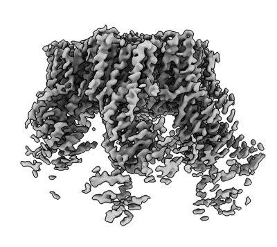
- EMDB-31988: Cryo-EM structure of the hexameric plasma membrane H+-ATPase in t... -
+ Open data

- Basic information
| Entry | Database: EMDB / ID: EMD-31988 | |||||||||
|---|---|---|---|---|---|---|---|---|---|---|
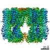
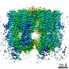
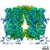
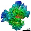
| Title | Cryo-EM structure of the hexameric plasma membrane H+-ATPase in the active state (pH 6.0, BeF3-, conformation 1, C1 symmetry) | |||||||||
 Map data Map data | ||||||||||
 Sample Sample |
| |||||||||
 Keywords Keywords | P type ATPase / plasma membrane H+-ATPase / hexamer / TRANSLOCASE | |||||||||
| Function / homology |  Function and homology information Function and homology informationP-type H+-exporting transporter / eisosome / proton export across plasma membrane / proteasome storage granule assembly / P-type proton-exporting transporter activity / positive regulation of TORC1 signaling / proton transmembrane transport / regulation of intracellular pH / transmembrane transport / membrane raft ...P-type H+-exporting transporter / eisosome / proton export across plasma membrane / proteasome storage granule assembly / P-type proton-exporting transporter activity / positive regulation of TORC1 signaling / proton transmembrane transport / regulation of intracellular pH / transmembrane transport / membrane raft / ATP hydrolysis activity / mitochondrion / ATP binding / metal ion binding / plasma membrane / cytosol Similarity search - Function | |||||||||
| Biological species |  | |||||||||
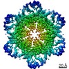
| Method | single particle reconstruction / cryo EM / Resolution: 3.8 Å | |||||||||
 Authors Authors | Zhao P / Zhao C | |||||||||
| Funding support |  China, 1 items China, 1 items
| |||||||||
 Citation Citation | Journal: Nat Commun / Year: 2021 Title: Structure and activation mechanism of the hexameric plasma membrane H-ATPase. Authors: Peng Zhao / Chaoran Zhao / Dandan Chen / Caihong Yun / Huilin Li / Lin Bai /  Abstract: The S. cerevisiae plasma membrane H-ATPase, Pma1, is a P3A-type ATPase and the primary protein component of the membrane compartment of Pma1 (MCP). Like other plasma membrane H-ATPases, Pma1 ...The S. cerevisiae plasma membrane H-ATPase, Pma1, is a P3A-type ATPase and the primary protein component of the membrane compartment of Pma1 (MCP). Like other plasma membrane H-ATPases, Pma1 assembles and functions as a hexamer, a property unique to this subfamily among the larger family of P-type ATPases. It has been unclear how Pma1 organizes the yeast membrane into MCP microdomains, or why it is that Pma1 needs to assemble into a hexamer to establish the membrane electrochemical proton gradient. Here we report a high-resolution cryo-EM study of native Pma1 hexamers embedded in endogenous lipids. Remarkably, we found that the Pma1 hexamer encircles a liquid-crystalline membrane domain composed of 57 ordered lipid molecules. The Pma1-encircled lipid patch structure likely serves as the building block of the MCP. At pH 7.4, the carboxyl-terminal regulatory α-helix binds to the phosphorylation domains of two neighboring Pma1 subunits, locking the hexamer in the autoinhibited state. The regulatory helix becomes disordered at lower pH, leading to activation of the Pma1 hexamer. The activation process is accompanied by a 6.7 Å downward shift and a 40° rotation of transmembrane helices 1 and 2 that line the proton translocation path. The conformational changes have enabled us to propose a detailed mechanism for ATP-hydrolysis-driven proton pumping across the plasma membrane. Our structures will facilitate the development of antifungal drugs that target this essential protein. | |||||||||
| History |
|
- Structure visualization
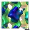
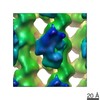
| Movie |
Movie viewer |
|---|---|
| Structure viewer | EM map: SurfViewMolmilJmol/JSmol |
| Supplemental images |
- Downloads & links
-EMDB archive
| Map data | emd_31988.map.gz | 12.2 MB | EMDB map data format | |
|---|---|---|---|---|
| Header (meta data) | emd-31988-v30.xmlemd-31988.xml | 13.4 KB 13.4 KB | Display Display | EMDB header |
| Images |  emd_31988.png emd_31988.png | 116 KB | ||
| Filedesc metadata | emd-31988.cif.gz | 6.3 KB | ||
| Archive directory |  http://ftp.pdbj.org/pub/emdb/structures/EMD-31988ftp://ftp.pdbj.org/pub/emdb/structures/EMD-31988 http://ftp.pdbj.org/pub/emdb/structures/EMD-31988ftp://ftp.pdbj.org/pub/emdb/structures/EMD-31988 | HTTPS FTP |
-Related structure data
| Related structure data | 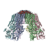 7vh6MC 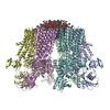 7vh5C M: atomic model generated by this map C: citing same article ( |
|---|---|
| Similar structure data |


-Links
| EMDB pages | EMDB (EBI/PDBe) / EMDataResource |
|---|---|
| Related items in Molecule of the Month |


-Map
| File | Download / File: emd_31988.map.gz / Format: CCP4 / Size: 103 MB / Type: IMAGE STORED AS FLOATING POINT NUMBER (4 BYTES) | ||||||||||||||||||||||||||||||||||||||||||||||||||||||||||||||||||||
|---|---|---|---|---|---|---|---|---|---|---|---|---|---|---|---|---|---|---|---|---|---|---|---|---|---|---|---|---|---|---|---|---|---|---|---|---|---|---|---|---|---|---|---|---|---|---|---|---|---|---|---|---|---|---|---|---|---|---|---|---|---|---|---|---|---|---|---|---|---|
| Projections & slices | Image control
Images are generated by Spider. | ||||||||||||||||||||||||||||||||||||||||||||||||||||||||||||||||||||
| Voxel size | X=Y=Z: 1.04 Å | ||||||||||||||||||||||||||||||||||||||||||||||||||||||||||||||||||||
| Density |
| ||||||||||||||||||||||||||||||||||||||||||||||||||||||||||||||||||||
| Symmetry | Space group: 1 | ||||||||||||||||||||||||||||||||||||||||||||||||||||||||||||||||||||
| Details | EMDB XML:
CCP4 map header:
| ||||||||||||||||||||||||||||||||||||||||||||||||||||||||||||||||||||
 Z (Sec.)
 Y (Row.)
 X (Col.)


-Supplemental data
- Sample components
-Entire : Plasma membrane ATPase 1
| Entire | Name: Plasma membrane ATPase 1 |
|---|---|
| Components |
|
-Supramolecule #1: Plasma membrane ATPase 1
| Supramolecule | Name: Plasma membrane ATPase 1 / type: complex / ID: 1 / Parent: 0 / Macromolecule list: #1 |
|---|---|
| Source (natural) | Organism: |
| Molecular weight | Theoretical: 99.6 kDa/nm |
-Macromolecule #1: Plasma membrane ATPase 1
| Macromolecule | Name: Plasma membrane ATPase 1 / type: protein_or_peptide / ID: 1 / Number of copies: 6 / Enantiomer: LEVO / EC number: P-type H+-exporting transporter |
|---|---|
| Source (natural) | Organism: Strain: ATCC 204508 / S288c |
| Molecular weight | Theoretical: 99.714023 KDa |
| Recombinant expression | Organism: |
| Sequence | String: MTDTSSSSSS SSASSVSAHQ PTQEKPAKTY DDAASESSDD DDIDALIEEL QSNHGVDDED SDNDGPVAAG EARPVPEEYL QTDPSYGLT SDEVLKRRKK YGLNQMADEK ESLVVKFVMF FVGPIQFVME AAAILAAGLS DWVDFGVICG LLMLNAGVGF V QEFQAGSI ...String: MTDTSSSSSS SSASSVSAHQ PTQEKPAKTY DDAASESSDD DDIDALIEEL QSNHGVDDED SDNDGPVAAG EARPVPEEYL QTDPSYGLT SDEVLKRRKK YGLNQMADEK ESLVVKFVMF FVGPIQFVME AAAILAAGLS DWVDFGVICG LLMLNAGVGF V QEFQAGSI VDELKKTLAN TAVVIRDGQL VEIPANEVVP GDILQLEDGT VIPTDGRIVT EDCFLQIDQS AITGESLAVD KH YGDQTFS SSTVKRGEGF MVVTATGDNT FVGRAAALVN KAAGGQGHFT EVLNGIGIIL LVLVIATLLL VWTACFYRTN GIV RILRYT LGITIIGVPV GLPAVVTTTM AVGAAYLAKK QAIVQKLSAI ESLAGVEILC SDKTGTLTKN KLSLHEPYTV EGVS PDDLM LTACLAASRK KKGLDAIDKA FLKSLKQYPK AKDALTKYKV LEFHPFDPVS KKVTAVVESP EGERIVCVKG APLFV LKTV EEDHPIPEDV HENYENKVAE LASRGFRALG VARKRGEGHW EILGVMPCMD PPRDDTAQTV SEARHLGLRV KMLTGD AVG IAKETCRQLG LGTNIYNAER LGLGGGGDMP GSELADFVEN ADGFAEVFPQ HKYRVVEILQ NRGYLVAMTG DGVNDAP SL KKADTGIAVE GATDAARSAA DIVFLAPGLS AIIDALKTSR QIFHRMYSYV VYRIALSLHL EIFLGLWIAI LDNSLDID L IVFIAIFADV ATLAIAYDNA PYSPKPVKWN LPRLWGMSII LGIVLAIGSW ITLTTMFLPK GGIIQNFGAM NGIMFLQIS LTENWLIFIT RAAGPFWSSI PSWQLAGAVF AVDIIATMFT LFGWWSENWT DIVTVVRVWI WSIGIFCVLG GFYYEMSTSE AFDRLMNGK PMKEKKSTRS VEDFMAAMQR VSTQHEKET UniProtKB: Plasma membrane ATPase 1 |
-Macromolecule #2: BERYLLIUM TRIFLUORIDE ION
| Macromolecule | Name: BERYLLIUM TRIFLUORIDE ION / type: ligand / ID: 2 / Number of copies: 6 / Formula: BEF |
|---|---|
| Molecular weight | Theoretical: 66.007 Da |
| Chemical component information |  ChemComp-BEF: |
-Macromolecule #3: (2S)-3-(hexadecanoyloxy)-2-[(9Z)-octadec-9-enoyloxy]propyl 2-(tri...
| Macromolecule | Name: (2S)-3-(hexadecanoyloxy)-2-[(9Z)-octadec-9-enoyloxy]propyl 2-(trimethylammonio)ethyl phosphate type: ligand / ID: 3 / Number of copies: 57 / Formula: POV |
|---|---|
| Molecular weight | Theoretical: 760.076 Da |
| Chemical component information |  ChemComp-POV: |
-Experimental details
-Structure determination
| Method | cryo EM |
|---|---|
 Processing Processing | single particle reconstruction |
| Aggregation state | particle |
-Sample preparation
| Buffer | pH: 7.4 |
|---|---|
| Vitrification | Cryogen name: ETHANE |
- Electron microscopy
| Microscope | FEI TITAN KRIOS |
|---|---|
| Image recording | Film or detector model: GATAN K2 SUMMIT (4k x 4k) / Average electron dose: 1.6 e/Å2 |
| Electron beam | Acceleration voltage: 300 kV / Electron source:  FIELD EMISSION GUN FIELD EMISSION GUN |
| Electron optics | Illumination mode: FLOOD BEAM / Imaging mode: BRIGHT FIELD |
| Experimental equipment |  Model: Titan Krios / Image courtesy: FEI Company |
