[English] 日本語
Yorodumi
- EMDB-31991: Cryo-EM structure of the hexameric plasma membrane H+-ATPase in t... -

+
Open data


ID or keywords:

Loading...

-
Basic information

Entry
Database: EMDB / ID: EMD-31991
TitleCryo-EM structure of the hexameric plasma membrane H+-ATPase in the autoinhibited state (pH 6.0, C1 symmetry)
Map data
Sample
  • Complex: Plasma membrane ATPase 1
    • Protein or peptide: Pma1
Function / homology
Function and homology information


eisosome / P-type H+-exporting transporter / proton export across plasma membrane / P-type ion transporter activity / P-type proton-exporting transporter activity / proteasome storage granule assembly / positive regulation of TORC1 signaling / proton transmembrane transport / monoatomic ion transmembrane transport / regulation of intracellular pH ...eisosome / P-type H+-exporting transporter / proton export across plasma membrane / P-type ion transporter activity / P-type proton-exporting transporter activity / proteasome storage granule assembly / positive regulation of TORC1 signaling / proton transmembrane transport / monoatomic ion transmembrane transport / regulation of intracellular pH / transmembrane transport / membrane => GO:0016020 / membrane raft / ATP hydrolysis activity / mitochondrion / ATP binding / metal ion binding / plasma membrane / cytosol
Similarity search - Function
P-type ATPase, subfamily IIIA / Cation transporter/ATPase, N-terminus / Cation-transporting P-type ATPase, N-terminal / Cation transporter/ATPase, N-terminus / P-type ATPase, haloacid dehalogenase domain / P-type ATPase, phosphorylation site / P-type ATPase, cytoplasmic domain N / E1-E2 ATPases phosphorylation site. / P-type ATPase, A domain superfamily / P-type ATPase ...P-type ATPase, subfamily IIIA / Cation transporter/ATPase, N-terminus / Cation-transporting P-type ATPase, N-terminal / Cation transporter/ATPase, N-terminus / P-type ATPase, haloacid dehalogenase domain / P-type ATPase, phosphorylation site / P-type ATPase, cytoplasmic domain N / E1-E2 ATPases phosphorylation site. / P-type ATPase, A domain superfamily / P-type ATPase / P-type ATPase, transmembrane domain superfamily / HAD superfamily / HAD-like superfamily
Similarity search - Domain/homology
Plasma membrane ATPase 1
Similarity search - Component
Biological speciesSaccharomyces cerevisiae (brewer's yeast)
Methodsingle particle reconstruction / cryo EM / Resolution: 3.8 Å
AuthorsZhao P / Zhao C / Chen D / Yun C / Li H / Bai L
Funding support China, 1 items
OrganizationGrant numberCountry
National Natural Science Foundation of China (NSFC)32171212 China
Citation
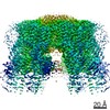
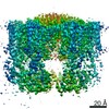
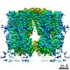
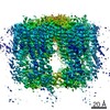
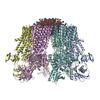
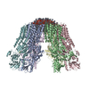
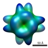
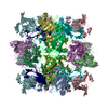
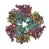
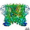
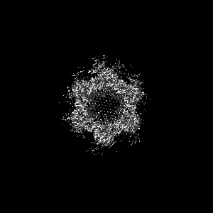
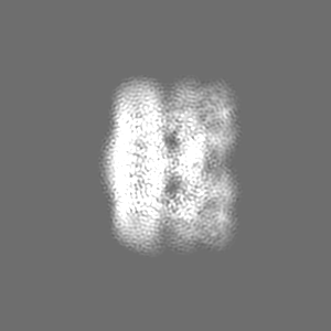
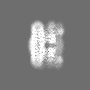
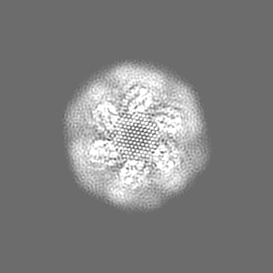
Journal: Nat Commun / Year: 2021
Title: Structure and activation mechanism of the hexameric plasma membrane H-ATPase.
Authors: Peng Zhao / Chaoran Zhao / Dandan Chen / Caihong Yun / Huilin Li / Lin Bai /
Abstract: The S. cerevisiae plasma membrane H-ATPase, Pma1, is a P3A-type ATPase and the primary protein component of the membrane compartment of Pma1 (MCP). Like other plasma membrane H-ATPases, Pma1 ...The S. cerevisiae plasma membrane H-ATPase, Pma1, is a P3A-type ATPase and the primary protein component of the membrane compartment of Pma1 (MCP). Like other plasma membrane H-ATPases, Pma1 assembles and functions as a hexamer, a property unique to this subfamily among the larger family of P-type ATPases. It has been unclear how Pma1 organizes the yeast membrane into MCP microdomains, or why it is that Pma1 needs to assemble into a hexamer to establish the membrane electrochemical proton gradient. Here we report a high-resolution cryo-EM study of native Pma1 hexamers embedded in endogenous lipids. Remarkably, we found that the Pma1 hexamer encircles a liquid-crystalline membrane domain composed of 57 ordered lipid molecules. The Pma1-encircled lipid patch structure likely serves as the building block of the MCP. At pH 7.4, the carboxyl-terminal regulatory α-helix binds to the phosphorylation domains of two neighboring Pma1 subunits, locking the hexamer in the autoinhibited state. The regulatory helix becomes disordered at lower pH, leading to activation of the Pma1 hexamer. The activation process is accompanied by a 6.7 Å downward shift and a 40° rotation of transmembrane helices 1 and 2 that line the proton translocation path. The conformational changes have enabled us to propose a detailed mechanism for ATP-hydrolysis-driven proton pumping across the plasma membrane. Our structures will facilitate the development of antifungal drugs that target this essential protein.
History
DepositionSep 21, 2021-
Header (metadata) releaseNov 17, 2021-
Map releaseNov 17, 2021-
UpdateNov 17, 2021-
Current statusNov 17, 2021Processing site: PDBj / Status: Released

-
Structure visualization
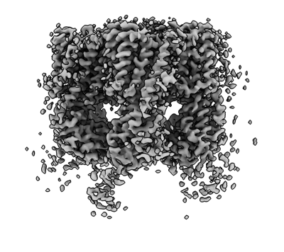
Movie
  • Surface view with section colored by density value
  • Surface level: 0.02
  • Imaged by UCSF Chimera
  • Download
  • Surface view colored by cylindrical radius
  • Surface level: 0.02
  • Imaged by UCSF Chimera
  • Download
Movie viewer
Structure viewerEM map:
SurfViewMolmilJmol/JSmol
Supplemental images

Downloads & links

-
Map

FileDownload / File: emd_31991.map.gz / Format: CCP4 / Size: 103 MB / Type: IMAGE STORED AS FLOATING POINT NUMBER (4 BYTES)
Voxel sizeX=Y=Z: 1.04 Å
Density
Contour LevelBy AUTHOR: 0.02 / Movie #1: 0.02
Minimum - Maximum-0.042312548 - 0.07120282
Average (Standard dev.)0.0003440812 (±0.002596157)
SymmetrySpace group: 1
Details

EMDB XML:

Map geometry
Axis orderXYZ
Origin000
Dimensions300300300
Spacing300300300
CellA=B=C: 312.0 Å
α=β=γ: 90.0 °

CCP4 map header:

modeImage stored as Reals
Å/pix. X/Y/Z1.041.041.04
M x/y/z300300300
origin x/y/z0.0000.0000.000
length x/y/z312.000312.000312.000
α/β/γ90.00090.00090.000
start NX/NY/NZ000
NX/NY/NZ256256256
MAP C/R/S123
start NC/NR/NS000
NC/NR/NS300300300
D min/max/mean-0.0420.0710.000

-
Supplemental data

-
Sample components

-
Entire : Plasma membrane ATPase 1

EntireName: Plasma membrane ATPase 1
Components
  • Complex: Plasma membrane ATPase 1
    • Protein or peptide: Pma1

-
Supramolecule #1: Plasma membrane ATPase 1

SupramoleculeName: Plasma membrane ATPase 1 / type: complex / ID: 1 / Parent: 0 / Macromolecule list: all
Source (natural)Organism: Saccharomyces cerevisiae (brewer's yeast)
Molecular weightExperimental: 99.6 kDa/nm

-
Macromolecule #1: Pma1

MacromoleculeName: Pma1 / type: protein_or_peptide / ID: 1 / Enantiomer: LEVO
Source (natural)Organism: Saccharomyces cerevisiae (brewer's yeast)
SequenceString: MTDTSSSSSS SSASSVSAHQ PTQEKPAKTY DDAASESSDD DDIDALIEEL QSNHGVDDED SDNDGPVAA GEARPVPEEY LQTDPSYGLT SDEVLKRRKK YGLNQMADEK ESLVVKFVMF F VGPIQFVM EAAAILAAGL SDWVDFGVIC GLLMLNAGVG FVQEFQAGSI ...String:
MTDTSSSSSS SSASSVSAHQ PTQEKPAKTY DDAASESSDD DDIDALIEEL QSNHGVDDED SDNDGPVAA GEARPVPEEY LQTDPSYGLT SDEVLKRRKK YGLNQMADEK ESLVVKFVMF F VGPIQFVM EAAAILAAGL SDWVDFGVIC GLLMLNAGVG FVQEFQAGSI VDELKKTLAN TA VVIRDGQ LVEIPANEVV PGDILQLEDG TVIPTDGRIV TEDCFLQIDQ SAITGESLAV DKH YGDQTF SSSTVKRGEG FMVVTATGDN TFVGRAAALV NKAAGGQGHF TEVLNGIGII LLVL VIATL LLVWTACFYR TNGIVRILRY TLGITIIGVP VGLPAVVTTT MAVGAAYLAK KQAIV QKLS AIESLAGVEI LCSDKTGTLT KNKLSLHEPY TVEGVSPDDL MLTACLAASR KKKGLD AID KAFLKSLKQY PKAKDALTKY KVLEFHPFDP VSKKVTAVVE SPEGERIVCV KGAPLFV LK TVEEDHPIPE DVHENYENKV AELASRGFRA LGVARKRGEG HWEILGVMPC MDPPRDDT A QTVSEARHLG LRVKMLTGDA VGIAKETCRQ LGLGTNIYNA ERLGLGGGGD MPGSELADF VENADGFAEV FPQHKYRVVE ILQNRGYLVA MTGDGVNDAP SLKKADTGIA VEGATDAARS AADIVFLAP GLSAIIDALK TSRQIFHRMY SYVVYRIALS LHLEIFLGLW IAILDNSLDI D LIVFIAIF ADVATLAIAY DNAPYSPKPV KWNLPRLWGM SIILGIVLAI GSWITLTTMF LP KGGIIQN FGAMNGIMFL QISLTENWLI FITRAAGPFW SSIPSWQLAG AVFAVDIIAT MFT LFGWWS ENWTDIVTVV RVWIWSIGIF CVLGGFYYEM STSEAFDRLM NGKPMKEKKS TRSV EDFMA AMQRVSTQHE KET

-
Experimental details

-
Structure determination

Methodcryo EM
Processingsingle particle reconstruction
Aggregation stateparticle

-
Sample preparation

BufferpH: 7.4
VitrificationCryogen name: ETHANE

-
Electron microscopy

MicroscopeFEI TITAN KRIOS
Electron beamAcceleration voltage: 300 kV / Electron source: FIELD EMISSION GUN
Electron opticsIllumination mode: FLOOD BEAM / Imaging mode: BRIGHT FIELDBright-field microscopy
Image recordingFilm or detector model: GATAN K2 SUMMIT (4k x 4k) / Average electron dose: 1.6 e/Å2
Experimental equipment
Model: Titan Krios / Image courtesy: FEI Company

-
Image processing

Initial angle assignmentType: ANGULAR RECONSTITUTION
Final angle assignmentType: ANGULAR RECONSTITUTION
Final reconstructionResolution.type: BY AUTHOR / Resolution: 3.8 Å / Resolution method: FSC 0.143 CUT-OFF / Number images used: 493148

+
About Yorodumi

-
News

-
Feb 9, 2022. New format data for meta-information of EMDB entries

New format data for meta-information of EMDB entries

  • Version 3 of the EMDB header file is now the official format.
  • The previous official version 1.9 will be removed from the archive.

Related info.:EMDB header

External links:wwPDB to switch to version 3 of the EMDB data model

-
Aug 12, 2020. Covid-19 info

Covid-19 info

URL: https://pdbj.org/emnavi/covid19.php

New page: Covid-19 featured information page in EM Navigator.

Related info.:Covid-19 info / Mar 5, 2020. Novel coronavirus structure data

+
Mar 5, 2020. Novel coronavirus structure data

Novel coronavirus structure data

Related info.:Yorodumi Speices / Aug 12, 2020. Covid-19 info

External links:COVID-19 featured content - PDBj / Molecule of the Month (242):Coronavirus Proteases

+
Jan 31, 2019. EMDB accession codes are about to change! (news from PDBe EMDB page)

EMDB accession codes are about to change! (news from PDBe EMDB page)

  • The allocation of 4 digits for EMDB accession codes will soon come to an end. Whilst these codes will remain in use, new EMDB accession codes will include an additional digit and will expand incrementally as the available range of codes is exhausted. The current 4-digit format prefixed with “EMD-” (i.e. EMD-XXXX) will advance to a 5-digit format (i.e. EMD-XXXXX), and so on. It is currently estimated that the 4-digit codes will be depleted around Spring 2019, at which point the 5-digit format will come into force.
  • The EM Navigator/Yorodumi systems omit the EMD- prefix.

Related info.:Q: What is EMD? / ID/Accession-code notation in Yorodumi/EM Navigator

External links:EMDB Accession Codes are Changing Soon! / Contact to PDBj

+
Jul 12, 2017. Major update of PDB

Major update of PDB

  • wwPDB released updated PDB data conforming to the new PDBx/mmCIF dictionary.
  • This is a major update changing the version number from 4 to 5, and with Remediation, in which all the entries are updated.
  • In this update, many items about electron microscopy experimental information are reorganized (e.g. em_software).
  • Now, EM Navigator and Yorodumi are based on the updated data.

External links:wwPDB Remediation / Enriched Model Files Conforming to OneDep Data Standards Now Available in the PDB FTP Archive

-
Yorodumi

Thousand views of thousand structures

  • Yorodumi is a browser for structure data from EMDB, PDB, SASBDB, etc.
  • This page is also the successor to EM Navigator detail page, and also detail information page/front-end page for Omokage search.
  • The word "yorodu" (or yorozu) is an old Japanese word meaning "ten thousand". "mi" (miru) is to see.

Related info.:EMDB / PDB / SASBDB / Comparison of 3 databanks / Yorodumi Search / Aug 31, 2016. New EM Navigator & Yorodumi / Yorodumi Papers / Jmol/JSmol / Function and homology information / Changes in new EM Navigator and Yorodumi

Read more