ムービー
ムービー コントローラー


+ データを開く

- 基本情報
基本情報
| 登録情報 | データベース: EMDB / ID: EMD-31953 | ||||||||||||
|---|---|---|---|---|---|---|---|---|---|---|---|---|---|
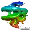
| タイトル | Cryo-EM structure of Vaccinia virus scaffolding protein D13 tubular assembly | ||||||||||||
 マップデータ マップデータ | Cryo-EM helical reconstruction of Vaccinia virus scaffold protein D13 tubular assembly. The map has been sharpened using Relion post-processing. The map has been density-normalized (mean=0, s.d=1). | ||||||||||||
 試料 試料 |
| ||||||||||||
| 生物種 |  Vaccinia virus WR (ウイルス) Vaccinia virus WR (ウイルス) | ||||||||||||
| 手法 | らせん対称体再構成法 / クライオ電子顕微鏡法 / 解像度: 7.33 Å | ||||||||||||
 データ登録者 データ登録者 | Hyun J / Matsunami H / Kim TG / Wolf M | ||||||||||||
| 資金援助 |  日本, 3件 日本, 3件
| ||||||||||||
 引用 引用 | ジャーナル: Nat Commun / 年: 2022 タイトル: Assembly mechanism of the pleomorphic immature poxvirus scaffold. 著者: Jaekyung Hyun / Hideyuki Matsunami / Tae Gyun Kim / Matthias Wolf /   要旨: In Vaccinia virus (VACV), the prototype poxvirus, scaffold protein D13 forms a honeycomb-like lattice on the viral membrane that results in formation of the pleomorphic immature virion (IV). The ...In Vaccinia virus (VACV), the prototype poxvirus, scaffold protein D13 forms a honeycomb-like lattice on the viral membrane that results in formation of the pleomorphic immature virion (IV). The structure of D13 is similar to those of major capsid proteins that readily form icosahedral capsids in nucleocytoplasmic large DNA viruses (NCLDVs). However, the detailed assembly mechanism of the nonicosahedral poxvirus scaffold has never been understood. Here we show the cryo-EM structures of the D13 trimer and scaffold intermediates produced in vitro. The structures reveal that the displacement of the short N-terminal α-helix is critical for initiation of D13 self-assembly. The continuous curvature of the IV is mediated by electrostatic interactions that induce torsion between trimers. The assembly mechanism explains the semiordered capsid-like arrangement of D13 that is distinct from icosahedral NCLDVs. Our structures explain how a single protein can self-assemble into different capsid morphologies and represent a local exception to the universal Caspar-Klug theory of quasi-equivalence. | ||||||||||||
| 履歴 |
|
- 構造の表示
構造の表示
| ムービー |
 ムービービューア ムービービューア |
|---|---|
| 構造ビューア | EMマップ: SurfViewMolmilJmol/JSmol |
| 添付画像 |

- ダウンロードとリンク
-EMDBアーカイブ
| マップデータ | emd_31953.map.gz | 84.7 MB | EMDBマップデータ形式 | |
|---|---|---|---|---|
| ヘッダ (付随情報) | emd-31953-v30.xmlemd-31953.xml | 20.1 KB 20.1 KB | 表示 表示 | EMDBヘッダ |
| FSC (解像度算出) | emd_31953_fsc.xml | 18.1 KB | 表示 | FSCデータファイル |
| 画像 |  emd_31953.png emd_31953.png | 201.1 KB | ||
| マスクデータ | emd_31953_msk_1.map | 512 MB | マスクマップ | |
| その他 | emd_31953_half_map_1.map.gzemd_31953_half_map_2.map.gz | 410.6 MB 410.7 MB | ||
| アーカイブディレクトリ |  http://ftp.pdbj.org/pub/emdb/structures/EMD-31953ftp://ftp.pdbj.org/pub/emdb/structures/EMD-31953 http://ftp.pdbj.org/pub/emdb/structures/EMD-31953ftp://ftp.pdbj.org/pub/emdb/structures/EMD-31953 | HTTPS FTP |
-検証レポート
| 文書・要旨 | emd_31953_validation.pdf.gz | 558.2 KB | 表示 | EMDB検証レポート |
|---|---|---|---|---|
| 文書・詳細版 | emd_31953_full_validation.pdf.gz | 557.7 KB | 表示 | |
| XML形式データ | emd_31953_validation.xml.gz | 26.3 KB | 表示 | |
| CIF形式データ | emd_31953_validation.cif.gz | 34.3 KB | 表示 | |
| アーカイブディレクトリ | https://ftp.pdbj.org/pub/emdb/validation_reports/EMD-31953ftp://ftp.pdbj.org/pub/emdb/validation_reports/EMD-31953 | HTTPS FTP |
-関連構造データ


-リンク
| EMDBのページ | EMDB (EBI/PDBe) / EMDataResource |
|---|
-マップ
| ファイル | ダウンロード / ファイル: emd_31953.map.gz / 形式: CCP4 / 大きさ: 512 MB / タイプ: IMAGE STORED AS FLOATING POINT NUMBER (4 BYTES) | ||||||||||||||||||||||||||||||||||||||||||||||||||||||||||||||||||||
|---|---|---|---|---|---|---|---|---|---|---|---|---|---|---|---|---|---|---|---|---|---|---|---|---|---|---|---|---|---|---|---|---|---|---|---|---|---|---|---|---|---|---|---|---|---|---|---|---|---|---|---|---|---|---|---|---|---|---|---|---|---|---|---|---|---|---|---|---|---|
| 注釈 | Cryo-EM helical reconstruction of Vaccinia virus scaffold protein D13 tubular assembly. The map has been sharpened using Relion post-processing. The map has been density-normalized (mean=0, s.d=1). | ||||||||||||||||||||||||||||||||||||||||||||||||||||||||||||||||||||
| 投影像・断面図 | 画像のコントロール
画像は Spider により作成 | ||||||||||||||||||||||||||||||||||||||||||||||||||||||||||||||||||||
| ボクセルのサイズ | X=Y=Z: 2.8 Å | ||||||||||||||||||||||||||||||||||||||||||||||||||||||||||||||||||||
| 密度 |
| ||||||||||||||||||||||||||||||||||||||||||||||||||||||||||||||||||||
| 対称性 | 空間群: 1 | ||||||||||||||||||||||||||||||||||||||||||||||||||||||||||||||||||||
| 詳細 | EMDB XML:
CCP4マップ ヘッダ情報:
| ||||||||||||||||||||||||||||||||||||||||||||||||||||||||||||||||||||
 Z (Sec.)
 Y (Row.)
 X (Col.)


-添付データ
-マスク #1
| ファイル | emd_31953_msk_1.map | ||||||||||||
|---|---|---|---|---|---|---|---|---|---|---|---|---|---|
| 投影像・断面図 |
| ||||||||||||
| 密度ヒストグラム |


-ハーフマップ: Cryo-EM helical reconstruction of Vaccinia virus scaffold protein...
| ファイル | emd_31953_half_map_1.map | ||||||||||||
|---|---|---|---|---|---|---|---|---|---|---|---|---|---|
| 注釈 | Cryo-EM helical reconstruction of Vaccinia virus scaffold protein D13 tubular assembly (half map 1). | ||||||||||||
| 投影像・断面図 |
| ||||||||||||
| 密度ヒストグラム |


-ハーフマップ: Cryo-EM helical reconstruction of Vaccinia virus scaffold protein...
| ファイル | emd_31953_half_map_2.map | ||||||||||||
|---|---|---|---|---|---|---|---|---|---|---|---|---|---|
| 注釈 | Cryo-EM helical reconstruction of Vaccinia virus scaffold protein D13 tubular assembly (half map 2). | ||||||||||||
| 投影像・断面図 |
| ||||||||||||
| 密度ヒストグラム |


- 試料の構成要素
-全体 : Vaccinia virus scaffolding protein D13 with N-terminal 17 residue...
| 全体 | 名称: Vaccinia virus scaffolding protein D13 with N-terminal 17 residue truncation, in its tubular assembly |
|---|---|
| 要素 |
|
-超分子 #1: Vaccinia virus scaffolding protein D13 with N-terminal 17 residue...
| 超分子 | 名称: Vaccinia virus scaffolding protein D13 with N-terminal 17 residue truncation, in its tubular assembly タイプ: complex / ID: 1 / 親要素: 0 / 含まれる分子: all 詳細: Recombinant D13 was expressed with N-terminal polyhistidine-tag (His-tag) using bacterial expression system. The protein was purified using metal affinity chromatography. His-tag was removed ...詳細: Recombinant D13 was expressed with N-terminal polyhistidine-tag (His-tag) using bacterial expression system. The protein was purified using metal affinity chromatography. His-tag was removed by proteolysis and the protein was further purified using size exclusion chromatography. The final purified protein was trimeric. The protein was assembled into tubes in low salt buffer. |
|---|---|
| 由来(天然) | 生物種: Vaccinia virus WR (ウイルス) |
| 組換発現 | 生物種:  |
| 分子量 | 理論値: 54 kDa/nm |
-分子 #1: Vaccinia virus scaffolding protein D13 with N-terminal 17 residue...
| 分子 | 名称: Vaccinia virus scaffolding protein D13 with N-terminal 17 residue truncation, in its tubular assembly タイプ: protein_or_peptide / ID: 1 / 光学異性体: LEVO |
|---|---|
| 由来(天然) | 生物種: Vaccinia virus WR (ウイルス) |
| 組換発現 | 生物種: |
| 配列 | 文字列: RSNVFAVDSQ IPTLYMPQYI SLSGVMTNDG PDNQAIASFE IRDQYITALN HLVLSLELPE VK GMGRFGY VPYVGYKCIN HVSISSCNGV IWEIEGEELY NNCINNTIAL KHSGYSSELN DISIGLTPND TIKEPSTVYV YIK TPFDVE DTFSSLKLSD SKITVTVTFN ...文字列: RSNVFAVDSQ IPTLYMPQYI SLSGVMTNDG PDNQAIASFE IRDQYITALN HLVLSLELPE VK GMGRFGY VPYVGYKCIN HVSISSCNGV IWEIEGEELY NNCINNTIAL KHSGYSSELN DISIGLTPND TIKEPSTVYV YIK TPFDVE DTFSSLKLSD SKITVTVTFN PVSDIVIRDS SFDFETFNKE FVYVPELSFI GYMVKNVQIK PSFIEKPRRV IGQI NQPTA TVTEVHAATS LSVYTKPYYG NTDNKFISYP GYSQDEKDYI DAYVSRLLDD LVIVSDGPPT GYPESAEIVE VPEDG IVSI QDADVYVKID NVPDNMSVYL HTNLLMFGTR KNSFIYNISK KFSAITGTYS DATKRTIFAH ISHSINIIDT SIPVSL WTS QRNVYNGDNR SAESKAKDLF INDPFIKGID FKNKTDIISR LEVRFGNDVL YSENGPISRI YNELLTKSNN GTRTLTF NF TPKIFFRPTT ITANVSRGKD KLSVRVVYST MDVNHPIYYV QKQLVVVCND LYKVSYDQGV SITKIMG |
-実験情報
-構造解析
| 手法 | クライオ電子顕微鏡法 |
|---|---|
 解析 解析 | らせん対称体再構成法 |
| 試料の集合状態 | particle |
-試料調製
| 濃度 | 0.12 mg/mL | ||||||||||||
|---|---|---|---|---|---|---|---|---|---|---|---|---|---|
| 緩衝液 | pH: 8 構成要素:
| ||||||||||||
| グリッド | モデル: Quantifoil R2/2 / 材質: COPPER / メッシュ: 300 / 支持フィルム - 材質: CARBON / 支持フィルム - トポロジー: HOLEY / 前処理 - タイプ: PLASMA CLEANING | ||||||||||||
| 凍結 | 凍結剤: ETHANE-PROPANE / チャンバー内湿度: 90 % / チャンバー内温度: 277 K / 装置: FEI VITROBOT MARK IV 詳細: 3 microliter sample volume was loaded onto a holey grid with additional graphene oxide film. 10 sec waiting time, 5 sec blotting time and blot force 0, no delay time were applied before plunging.. |
- 電子顕微鏡法
| 顕微鏡 | FEI TITAN KRIOS |
|---|---|
| 撮影 | フィルム・検出器のモデル: GATAN K2 QUANTUM (4k x 4k) 検出モード: COUNTING / デジタル化 - サイズ - 横: 4096 pixel / デジタル化 - サイズ - 縦: 4096 pixel / デジタル化 - サンプリング間隔: 5.0 µm / 撮影したグリッド数: 2 / 実像数: 7621 / 平均露光時間: 10.0 sec. / 平均電子線量: 50.0 e/Å2 |
| 電子線 | 加速電圧: 300 kV / 電子線源:  FIELD EMISSION GUN FIELD EMISSION GUN |
| 電子光学系 | C2レンズ絞り径: 70.0 µm / 照射モード: FLOOD BEAM / 撮影モード: BRIGHT FIELD / Cs: 2.7 mm / 最大 デフォーカス(公称値): 2.5 µm / 最小 デフォーカス(公称値): 0.5 µm / 倍率(公称値): 105000 |
| 試料ステージ | 試料ホルダーモデル: FEI TITAN KRIOS AUTOGRID HOLDER ホルダー冷却材: NITROGEN |
| 実験機器 |  モデル: Titan Krios / 画像提供: FEI Company |
+画像解析

-原子モデル構築 1
| 初期モデル | PDB ID: |
|---|---|
| 精密化 | 空間: REAL / プロトコル: RIGID BODY FIT |