Movie
Movie Controller

[English] 日本語
 Yorodumi
- PDB-7vfe: Cryo-EM structure of Vaccinia virus scaffolding protein D13 with ... -
+ Open data

- Basic information
| Entry | Database: PDB / ID: 7vfe | ||||||||||||
|---|---|---|---|---|---|---|---|---|---|---|---|---|---|
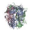
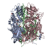
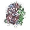
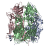
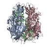
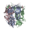
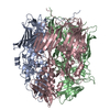
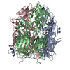
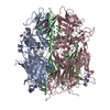
| Title | Cryo-EM structure of Vaccinia virus scaffolding protein D13 with N-terminal polyhistidine tag | ||||||||||||
 Components Components | Scaffold protein D13 | ||||||||||||
 Keywords Keywords | VIRAL PROTEIN / Scaffold / capsid / double-jelly-roll | ||||||||||||
| Function / homology | Poxvirus rifampicin-resistance / Poxvirus rifampicin resistance protein / response to antibiotic / membrane / identical protein binding / Scaffold protein OPG125 Function and homology information Function and homology information | ||||||||||||
| Biological species |  Vaccinia virus Vaccinia virus | ||||||||||||
| Method | ELECTRON MICROSCOPY / single particle reconstruction / cryo EM / Resolution: 2.63 Å | ||||||||||||
 Authors Authors | Wolf, M. / Hyun, J. / Matsunami, H. / Kim, T.G. | ||||||||||||
| Funding support |  Japan, 3items Japan, 3items
| ||||||||||||
 Citation Citation | Journal: Nat Commun / Year: 2022 Title: Assembly mechanism of the pleomorphic immature poxvirus scaffold. Authors: Jaekyung Hyun / Hideyuki Matsunami / Tae Gyun Kim / Matthias Wolf /   Abstract: In Vaccinia virus (VACV), the prototype poxvirus, scaffold protein D13 forms a honeycomb-like lattice on the viral membrane that results in formation of the pleomorphic immature virion (IV). The ...In Vaccinia virus (VACV), the prototype poxvirus, scaffold protein D13 forms a honeycomb-like lattice on the viral membrane that results in formation of the pleomorphic immature virion (IV). The structure of D13 is similar to those of major capsid proteins that readily form icosahedral capsids in nucleocytoplasmic large DNA viruses (NCLDVs). However, the detailed assembly mechanism of the nonicosahedral poxvirus scaffold has never been understood. Here we show the cryo-EM structures of the D13 trimer and scaffold intermediates produced in vitro. The structures reveal that the displacement of the short N-terminal α-helix is critical for initiation of D13 self-assembly. The continuous curvature of the IV is mediated by electrostatic interactions that induce torsion between trimers. The assembly mechanism explains the semiordered capsid-like arrangement of D13 that is distinct from icosahedral NCLDVs. Our structures explain how a single protein can self-assemble into different capsid morphologies and represent a local exception to the universal Caspar-Klug theory of quasi-equivalence. | ||||||||||||
| History |
|
- Structure visualization
| Movie |
Movie viewer |
|---|---|
| Structure viewer | Molecule: MolmilJmol/JSmol |
- Downloads & links
-Download
| PDBx/mmCIF format | 7vfe.cif.gz | 280.8 KB | Display | PDBx/mmCIF format |
|---|---|---|---|---|
| PDB format | pdb7vfe.ent.gz | 227.3 KB | Display | PDB format |
| PDBx/mmJSON format | 7vfe.json.gz | Tree view | PDBx/mmJSON format | |
| Others |  Other downloads Other downloads |
-Validation report
| Arichive directory | https://data.pdbj.org/pub/pdb/validation_reports/vf/7vfeftp://data.pdbj.org/pub/pdb/validation_reports/vf/7vfe | HTTPS FTP |
|---|
-Related structure data
| Related structure data |  31950MC  7vfdC  7vffC  7vfgC  7vfhC M: map data used to model this data C: citing same article ( |
|---|---|
| Similar structure data |


-Links
 PDBj
PDBj- Assembly
| Deposited unit | 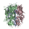
|
|---|---|
| 1 |
|
-Components
| #1: Protein | Mass: 64162.930 Da / Num. of mol.: 3 Source method: isolated from a genetically manipulated source Source: (gene. exp.) Vaccinia virus (strain Western Reserve)Strain: Western Reserve / Gene: VACWR118, D13L / Production host:  |
|---|
-Experimental details
-Experiment
| Experiment | Method: ELECTRON MICROSCOPY |
|---|---|
| EM experiment | Aggregation state: PARTICLE / 3D reconstruction method: single particle reconstruction |
- Sample preparation
| Component | Name: Vaccinia virus scaffolding protein D13 with N-terminal polyhistidine tag in its trimeric state Type: COMPLEX Details: Recombinant D13 was expressed with N-terminal polyhistidine-tag using bacterial expression system. The protein was purified using metal affinity chromatography and size exclusion ...Details: Recombinant D13 was expressed with N-terminal polyhistidine-tag using bacterial expression system. The protein was purified using metal affinity chromatography and size exclusion chromatography. The final purified protein was trimeric. Entity ID: all / Source: RECOMBINANT | ||||||||||||||||||||||||||||||
|---|---|---|---|---|---|---|---|---|---|---|---|---|---|---|---|---|---|---|---|---|---|---|---|---|---|---|---|---|---|---|---|
| Molecular weight | Value: 0.195 MDa / Experimental value: NO | ||||||||||||||||||||||||||||||
| Source (natural) | Organism: Vaccinia virus WR | ||||||||||||||||||||||||||||||
| Source (recombinant) | Organism: | ||||||||||||||||||||||||||||||
| Buffer solution | pH: 8 | ||||||||||||||||||||||||||||||
| Buffer component |
| ||||||||||||||||||||||||||||||
| Specimen | Conc.: 0.12 mg/ml / Embedding applied: NO / Shadowing applied: NO / Staining applied: NO / Vitrification applied: YES | ||||||||||||||||||||||||||||||
| Specimen support | Grid material: COPPER / Grid mesh size: 300 divisions/in. / Grid type: Quantifoil R1.2/1.3 | ||||||||||||||||||||||||||||||
| Vitrification | Instrument: FEI VITROBOT MARK IV / Cryogen name: ETHANE-PROPANE / Humidity: 90 % / Chamber temperature: 277 K Details: 3 microliter sample volume was loaded onto a holey grid with additional graphene oxide film. 10 sec waiting time, 5 sec blotting time and blot force 0, no delay time were applied before plunging. |
- Electron microscopy imaging
| Experimental equipment |  Model: Titan Krios / Image courtesy: FEI Company |
|---|---|
| Microscopy | Model: FEI TITAN KRIOS |
| Electron gun | Electron source:  FIELD EMISSION GUN / Accelerating voltage: 300 kV / Illumination mode: FLOOD BEAM FIELD EMISSION GUN / Accelerating voltage: 300 kV / Illumination mode: FLOOD BEAM |
| Electron lens | Mode: BRIGHT FIELD / Nominal magnification: 155000 X / Nominal defocus max: 1500 nm / Nominal defocus min: 500 nm / Cs: 2.7 mm / C2 aperture diameter: 70 µm / Alignment procedure: COMA FREE |
| Specimen holder | Cryogen: NITROGEN / Specimen holder model: FEI TITAN KRIOS AUTOGRID HOLDER |
| Image recording | Average exposure time: 31 sec. / Electron dose: 50 e/Å2 / Film or detector model: FEI FALCON III (4k x 4k) / Num. of grids imaged: 1 / Num. of real images: 1418 |
| Image scans | Sampling size: 14 µm / Width: 4096 / Height: 4096 |
- Processing
| Software | Name: PHENIX / Version: 1.18rc7_3834: / Classification: refinement | ||||||||||||||||||||||||||||||||||||||||||||||||||||||||||||
|---|---|---|---|---|---|---|---|---|---|---|---|---|---|---|---|---|---|---|---|---|---|---|---|---|---|---|---|---|---|---|---|---|---|---|---|---|---|---|---|---|---|---|---|---|---|---|---|---|---|---|---|---|---|---|---|---|---|---|---|---|---|
| EM software |
| ||||||||||||||||||||||||||||||||||||||||||||||||||||||||||||
| CTF correction | Type: PHASE FLIPPING AND AMPLITUDE CORRECTION | ||||||||||||||||||||||||||||||||||||||||||||||||||||||||||||
| Particle selection | Num. of particles selected: 284340 | ||||||||||||||||||||||||||||||||||||||||||||||||||||||||||||
| Symmetry | Point symmetry: C3 (3 fold cyclic) | ||||||||||||||||||||||||||||||||||||||||||||||||||||||||||||
| 3D reconstruction | Resolution: 2.63 Å / Resolution method: FSC 0.143 CUT-OFF / Num. of particles: 173354 / Algorithm: FOURIER SPACE / Num. of class averages: 1 / Symmetry type: POINT | ||||||||||||||||||||||||||||||||||||||||||||||||||||||||||||
| Atomic model building | Protocol: FLEXIBLE FIT / Space: REAL | ||||||||||||||||||||||||||||||||||||||||||||||||||||||||||||
| Atomic model building | PDB-ID: 6BEI Accession code: 6BEI / Source name: PDB / Type: experimental model | ||||||||||||||||||||||||||||||||||||||||||||||||||||||||||||
| Refine LS restraints |
|