 Movie
Movie Controller

[English] 日本語
 Yorodumi
- EMDB-31633: human Serine beta-lactamase-like protein LACTB truncation variant -
+ Open data

- Basic information
| Entry | Database: EMDB / ID: EMD-31633 | |||||||||
|---|---|---|---|---|---|---|---|---|---|---|
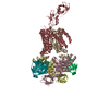
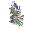
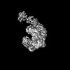
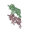
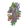
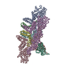
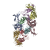
| Title | human Serine beta-lactamase-like protein LACTB truncation variant | |||||||||
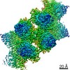
 Map data Map data | ||||||||||
 Sample Sample |
| |||||||||
 Keywords Keywords | mitochondrial intermembrane space protease / CYTOSOLIC PROTEIN / HYDROLASE | |||||||||
| Function / homology |  Function and homology information Function and homology informationHydrolases; Acting on peptide bonds (peptidases) / regulation of lipid metabolic process / lipid metabolic process / peptidase activity / mitochondrion / proteolysis / identical protein binding / cytosol Similarity search - Function | |||||||||
| Biological species |  Homo sapiens (human) Homo sapiens (human) | |||||||||
| Method | single particle reconstruction / cryo EM / Resolution: 3.08 Å | |||||||||
 Authors Authors | Zhang MH / Yang MJ | |||||||||
| Funding support |  China, 1 items China, 1 items
| |||||||||
 Citation Citation | Journal: Structure / Year: 2022 Title: Structural basis for the catalytic activity of filamentous human serine beta-lactamase-like protein LACTB. Authors: Minghui Zhang / Laixing Zhang / Runyu Guo / Chun Xiao / Jian Yin / Sensen Zhang / Maojun Yang / Abstract: Serine beta-lactamase-like protein (LACTB) is a mammalian mitochondrial serine protease that can specifically hydrolyze peptide bonds adjacent to aspartic acid residues and is structurally related to ...Serine beta-lactamase-like protein (LACTB) is a mammalian mitochondrial serine protease that can specifically hydrolyze peptide bonds adjacent to aspartic acid residues and is structurally related to prokaryotic penicillin-binding proteins. Here, we determined the cryoelectron microscopy structures of human LACTB (hLACTB) filaments from wild-type protein, a middle region deletion mutant, and in complex with the inhibitor Z-AAD-CMK at 3.0-, 3.1-, and 2.8-Å resolution, respectively. Structural analysis and activity assays revealed that three interfaces are required for the assembly of hLACTB filaments and that the formation of higher order helical structures facilitates its cleavage activity. Further structural and enzymatic analyses of middle region deletion constructs indicated that, while this region is necessary for substrate hydrolysis, it is not required for filament formation. Moreover, the inhibitor-bound structure showed that hLACTB may cleave peptide bonds adjacent to aspartic acid residues. These findings provide the structural basis underlying hLACTB catalytic activity. | |||||||||
| History |
|
- Structure visualization
| Movie |
Movie viewer |
|---|---|
| Structure viewer | EM map: SurfViewMolmilJmol/JSmol |
| Supplemental images |

- Downloads & links
-EMDB archive
| Map data | emd_31633.map.gz | 7.5 MB | EMDB map data format | |
|---|---|---|---|---|
| Header (meta data) | emd-31633-v30.xmlemd-31633.xml | 11.8 KB 11.8 KB | Display Display | EMDB header |
| Images | 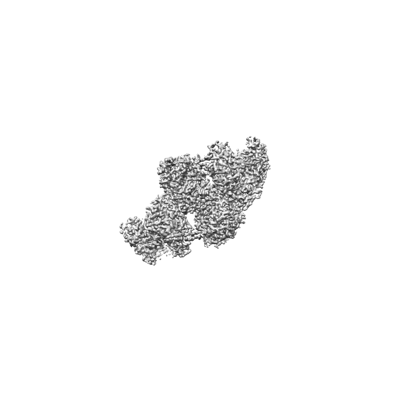 emd_31633.png emd_31633.png | 30.7 KB | ||
| Filedesc metadata | emd-31633.cif.gz | 5.6 KB | ||
| Archive directory |  http://ftp.pdbj.org/pub/emdb/structures/EMD-31633ftp://ftp.pdbj.org/pub/emdb/structures/EMD-31633 http://ftp.pdbj.org/pub/emdb/structures/EMD-31633ftp://ftp.pdbj.org/pub/emdb/structures/EMD-31633 | HTTPS FTP |
-Related structure data
| Related structure data | 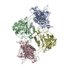 7v21MC  7v1yC 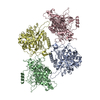 7v1zC M: atomic model generated by this map C: citing same article ( |
|---|---|
| Similar structure data |


-Links
| EMDB pages | EMDB (EBI/PDBe) / EMDataResource |
|---|---|
| Related items in Molecule of the Month |

-Map
| File | Download / File: emd_31633.map.gz / Format: CCP4 / Size: 83.7 MB / Type: IMAGE STORED AS FLOATING POINT NUMBER (4 BYTES) | ||||||||||||||||||||||||||||||||||||||||||||||||||||||||||||||||||||
|---|---|---|---|---|---|---|---|---|---|---|---|---|---|---|---|---|---|---|---|---|---|---|---|---|---|---|---|---|---|---|---|---|---|---|---|---|---|---|---|---|---|---|---|---|---|---|---|---|---|---|---|---|---|---|---|---|---|---|---|---|---|---|---|---|---|---|---|---|---|
| Projections & slices | Image control
Images are generated by Spider. | ||||||||||||||||||||||||||||||||||||||||||||||||||||||||||||||||||||
| Voxel size | X=Y=Z: 0.97 Å | ||||||||||||||||||||||||||||||||||||||||||||||||||||||||||||||||||||
| Density |
| ||||||||||||||||||||||||||||||||||||||||||||||||||||||||||||||||||||
| Symmetry | Space group: 1 | ||||||||||||||||||||||||||||||||||||||||||||||||||||||||||||||||||||
| Details | EMDB XML:
CCP4 map header:
| ||||||||||||||||||||||||||||||||||||||||||||||||||||||||||||||||||||
 Z (Sec.)
 Y (Row.)
 X (Col.)


-Supplemental data
- Sample components
-Entire : mitochondrial intermembrane space protease truncation
| Entire | Name: mitochondrial intermembrane space protease truncation |
|---|---|
| Components |
|
-Supramolecule #1: mitochondrial intermembrane space protease truncation
| Supramolecule | Name: mitochondrial intermembrane space protease truncation / type: organelle_or_cellular_component / ID: 1 / Parent: 0 / Macromolecule list: all |
|---|---|
| Source (natural) | Organism: Homo sapiens (human) |
-Macromolecule #1: Serine beta-lactamase-like protein LACTB, mitochondrial
| Macromolecule | Name: Serine beta-lactamase-like protein LACTB, mitochondrial type: protein_or_peptide / ID: 1 / Number of copies: 4 / Enantiomer: LEVO / EC number: Hydrolases; Acting on peptide bonds (peptidases) |
|---|---|
| Source (natural) | Organism: Homo sapiens (human) |
| Molecular weight | Theoretical: 47.029566 KDa |
| Recombinant expression | Organism:  |
| Sequence | String: GPAAPAQSPA APDPEASPLA EPPQEQSLAP WSPQTPAPPC SRCFARAIES SRDLLHRIKD EVGAPGIVVG VSVDGKEVWS EGLGYADVE NRVPCKPETV MRIASISKSL TMVALAKLWE AGKLDLDIPV QHYVPEFPEK EYEGEKVSVT TRLLISHLSG I RHYGELYL ...String: GPAAPAQSPA APDPEASPLA EPPQEQSLAP WSPQTPAPPC SRCFARAIES SRDLLHRIKD EVGAPGIVVG VSVDGKEVWS EGLGYADVE NRVPCKPETV MRIASISKSL TMVALAKLWE AGKLDLDIPV QHYVPEFPEK EYEGEKVSVT TRLLISHLSG I RHYGELYL REKFENSIES LRLFKNDPLF FKPGSQFLYS TFGYTLLAAI VERASGCKYL DYMQKIFHDL DMLTTVQEEN EP VIYNRAR FYVYNKKKRL VNTPYVDNSY KWAGGGFLST VGDLLKFGNA MLYGYQVGLF KNSNENLLPG YLKPETMVMM WTP VPNTEM SWDKEGKYAM AWGVVERKQT YGSCRKQRHY ASHTGGAVGA SSVLLVLPEE LDTETINNKV PPRGIIVSII CNMQ SVGLN STALKIALEF DKDRSD UniProtKB: Serine beta-lactamase-like protein LACTB, mitochondrial, Serine beta-lactamase-like protein LACTB, mitochondrial |
-Experimental details
-Structure determination
| Method | cryo EM |
|---|---|
 Processing Processing | single particle reconstruction |
| Aggregation state | particle |
-Sample preparation
| Buffer | pH: 7.4 |
|---|---|
| Vitrification | Cryogen name: ETHANE |
- Electron microscopy
| Microscope | FEI TITAN KRIOS |
|---|---|
| Image recording | Film or detector model: GATAN K3 (6k x 4k) / Average electron dose: 50.0 e/Å2 |
| Electron beam | Acceleration voltage: 300 kV / Electron source:  FIELD EMISSION GUN FIELD EMISSION GUN |
| Electron optics | Illumination mode: FLOOD BEAM / Imaging mode: BRIGHT FIELD |
| Experimental equipment |  Model: Titan Krios / Image courtesy: FEI Company |
