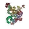
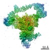
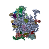
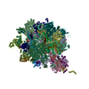
Journal: Nat Commun / Year: 2021 Title: Structural insight into the mechanism of energy transfer in cyanobacterial phycobilisomes. Authors: Lvqin Zheng / Zhenggao Zheng / Xiying Li / Guopeng Wang / Kun Zhang / Peijun Wei / Jindong Zhao / Ning Gao / Abstract: Phycobilisomes (PBS) are the major light-harvesting machineries for photosynthesis in cyanobacteria and red algae and they have a hierarchical structure of a core and peripheral rods, with both ...Phycobilisomes (PBS) are the major light-harvesting machineries for photosynthesis in cyanobacteria and red algae and they have a hierarchical structure of a core and peripheral rods, with both consisting of phycobiliproteins and linker proteins. Here we report the cryo-EM structures of PBS from two cyanobacterial species, Anabaena 7120 and Synechococcus 7002. Both PBS are hemidiscoidal in shape and share a common triangular core structure. While the Anabaena PBS has two additional hexamers in the core linked by the 4th linker domain of ApcE (L). The PBS structures predict that, compared with the PBS from red algae, the cyanobacterial PBS could have more direct routes for energy transfer to ApcD. Structure-based systematic mutagenesis analysis of the chromophore environment of ApcD and ApcF subunits reveals that aromatic residues are critical to excitation energy transfer (EET). The structures also suggest that the linker protein could actively participate in the process of EET in both rods and the cores. These results provide insights into the organization of chromophores and the mechanisms of EET within cyanobacterial PBS.
History
Deposition
Jun 26, 2021
-
Header (metadata) release
Oct 6, 2021
-
Map release
Oct 6, 2021
-
Update
Oct 6, 2021
-
Current status
Oct 6, 2021
Processing site: PDBj / Status: Released
-
Structure visualization
Movie
Surface view with section colored by density value
In the structure databanks used in Yorodumi, some data are registered as the other names, "COVID-19 virus" and "2019-nCoV". Here are the details of the virus and the list of structure data.
Jan 31, 2019. EMDB accession codes are about to change! (news from PDBe EMDB page)
EMDB accession codes are about to change! (news from PDBe EMDB page)
The allocation of 4 digits for EMDB accession codes will soon come to an end. Whilst these codes will remain in use, new EMDB accession codes will include an additional digit and will expand incrementally as the available range of codes is exhausted. The current 4-digit format prefixed with “EMD-” (i.e. EMD-XXXX) will advance to a 5-digit format (i.e. EMD-XXXXX), and so on. It is currently estimated that the 4-digit codes will be depleted around Spring 2019, at which point the 5-digit format will come into force.
The EM Navigator/Yorodumi systems omit the EMD- prefix.
Related info.:Q: What is EMD? / ID/Accession-code notation in Yorodumi/EM Navigator
Yorodumi is a browser for structure data from EMDB, PDB, SASBDB, etc.
This page is also the successor to EM Navigator detail page, and also detail information page/front-end page for Omokage search.
The word "yorodu" (or yorozu) is an old Japanese word meaning "ten thousand". "mi" (miru) is to see.
Related info.:EMDB / PDB / SASBDB / Comparison of 3 databanks / Yorodumi Search / Aug 31, 2016. New EM Navigator & Yorodumi / Yorodumi Papers / Jmol/JSmol / Function and homology information / Changes in new EM Navigator and Yorodumi
 Movie
Movie Controller

 Yorodumi
 Open data
 Basic information
 Map data
 Sample
 Function and homology information
 Synechococcus sp. PCC 7002 (bacteria)
 Authors
 Citation
 Structure visualization
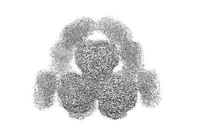
 Downloads & links
 emd_31483.png
 http://ftp.pdbj.org/pub/emdb/structures/EMD-31483


 Z (Sec.)
 Y (Row.)
 X (Col.)


 Sample components
 Processing
 Electron microscopy
 FIELD EMISSION GUN
