 Movie
Movie Controller


+ Open data

- Basic information
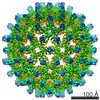
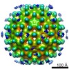
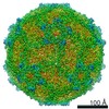
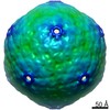
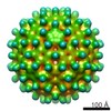
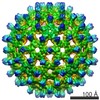
| Entry | Database: EMDB / ID: EMD-31234 | |||||||||
|---|---|---|---|---|---|---|---|---|---|---|
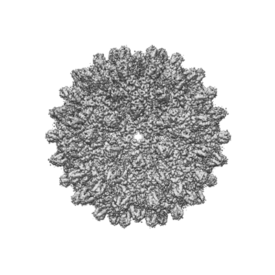
| Title | Engineered Hepatitis B virus core antigen T=4 | |||||||||
 Map data Map data | ||||||||||
 Sample Sample | Staphylococcus aureus != Hepatitis B virus adr/Japan/Nishioka/1983 Staphylococcus aureus
| |||||||||
 Keywords Keywords | cancer therapy / epidermal growth factor receptor 1 / affibody / VIRUS LIKE PARTICLE | |||||||||
| Function / homology |  Function and homology information Function and homology informationmicrotubule-dependent intracellular transport of viral material towards nucleus / IgG binding / T=4 icosahedral viral capsid / viral penetration into host nucleus / host cell / host cell cytoplasm / symbiont entry into host cell / structural molecule activity / DNA binding / RNA binding Similarity search - Function | |||||||||
| Biological species |  Hepatitis B virus genotype C subtype adr (strain Japan/adr4/1983) / Hepatitis B virus adr/Japan/Nishioka/1983 Hepatitis B virus genotype C subtype adr (strain Japan/adr4/1983) / Hepatitis B virus adr/Japan/Nishioka/1983 | |||||||||
| Method | single particle reconstruction / cryo EM / Resolution: 3.86 Å | |||||||||
 Authors Authors | Jeong H / Heo Y | |||||||||
 Citation Citation | Journal: Int J Mol Sci / Year: 2021 Title: Structural and Functional Characterizations of Cancer Targeting Nanoparticles Based on Hepatitis B Virus Capsid. Authors: Yunseok Heo / Hyeongseop Jeong / Youngki Yoo / Ji-Hye Yun / Bumhan Ryu / Young-Je Cha / Bo-Ram Lee / Ye-Eun Jeon / Jongmin Kim / Sojin Jeong / Eunji Jo / Jae-Sung Woo / Jeewon Lee / Hyun-Soo Cho / Weontae Lee /  Abstract: Cancer targeting nanoparticles have been extensively studied, but stable and applicable agents have yet to be developed. Here, we report stable nanoparticles based on hepatitis B core antigen (HBcAg) ...Cancer targeting nanoparticles have been extensively studied, but stable and applicable agents have yet to be developed. Here, we report stable nanoparticles based on hepatitis B core antigen (HBcAg) for cancer therapy. HBcAg monomers assemble into spherical capsids of 180 or 240 subunits. HBcAg was engineered to present an affibody for binding to human epidermal growth factor receptor 1 (EGFR) and to present histidine and tyrosine tags for binding to gold ions. The HBcAg engineered to present affibody and tags (HAF) bound specifically to EGFR and exterminated the EGFR-overexpressing adenocarcinomas under alternating magnetic field (AMF) after binding with gold ions. Using cryogenic electron microscopy (cryo-EM), we obtained the molecular structures of recombinant HAF and found that the overall structure of HAF was the same as that of HBcAg, except with the affibody on the spike. Therefore, HAF is viable for cancer therapy with the advantage of maintaining a stable capsid form. If the affibody in HAF is replaced with a specific sequence to bind to another targetable disease protein, the nanoparticles can be used for drug development over a wide spectrum. | |||||||||
| History |
|
- Structure visualization
| Movie |
Movie viewer |
|---|---|
| Structure viewer | EM map: SurfViewMolmilJmol/JSmol |
| Supplemental images |
- Downloads & links
-EMDB archive
| Map data | emd_31234.map.gz | 327.9 MB | EMDB map data format | |
|---|---|---|---|---|
| Header (meta data) | emd-31234-v30.xmlemd-31234.xml | 12.8 KB 12.8 KB | Display Display | EMDB header |
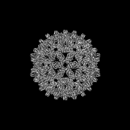
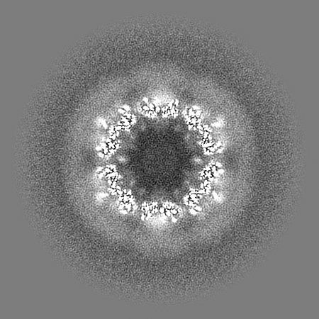
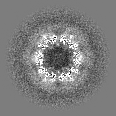
| Images |  emd_31234.png emd_31234.png | 118.4 KB | ||
| Filedesc metadata | emd-31234.cif.gz | 5.7 KB | ||
| Archive directory |  http://ftp.pdbj.org/pub/emdb/structures/EMD-31234ftp://ftp.pdbj.org/pub/emdb/structures/EMD-31234 http://ftp.pdbj.org/pub/emdb/structures/EMD-31234ftp://ftp.pdbj.org/pub/emdb/structures/EMD-31234 | HTTPS FTP |
-Related structure data
| Related structure data |  7ep6MC  7eoyC  7fdjC M: atomic model generated by this map C: citing same article ( |
|---|---|
| Similar structure data |

-Links
| EMDB pages | EMDB (EBI/PDBe) / EMDataResource |
|---|---|
| Related items in Molecule of the Month |


-Map
| File | Download / File: emd_31234.map.gz / Format: CCP4 / Size: 347.6 MB / Type: IMAGE STORED AS FLOATING POINT NUMBER (4 BYTES) | ||||||||||||||||||||||||||||||||||||||||||||||||||||||||||||
|---|---|---|---|---|---|---|---|---|---|---|---|---|---|---|---|---|---|---|---|---|---|---|---|---|---|---|---|---|---|---|---|---|---|---|---|---|---|---|---|---|---|---|---|---|---|---|---|---|---|---|---|---|---|---|---|---|---|---|---|---|---|
| Projections & slices | Image control
Images are generated by Spider. | ||||||||||||||||||||||||||||||||||||||||||||||||||||||||||||
| Voxel size | X=Y=Z: 1.4 Å | ||||||||||||||||||||||||||||||||||||||||||||||||||||||||||||
| Density |
| ||||||||||||||||||||||||||||||||||||||||||||||||||||||||||||
| Symmetry | Space group: 1 | ||||||||||||||||||||||||||||||||||||||||||||||||||||||||||||
| Details | EMDB XML:
CCP4 map header:
| ||||||||||||||||||||||||||||||||||||||||||||||||||||||||||||
 Z (Sec.)
 Y (Row.)
 X (Col.)


-Supplemental data
- Sample components
-Entire : Staphylococcus aureus
| Entire | Name:   Staphylococcus aureus (bacteria) Staphylococcus aureus (bacteria) |
|---|---|
| Components |
|
-Supramolecule #1: Hepatitis B virus adr/Japan/Nishioka/1983
| Supramolecule | Name: Hepatitis B virus adr/Japan/Nishioka/1983 / type: virus / ID: 1 / Parent: 0 / Macromolecule list: all / Details: virus core antigen / NCBI-ID: 482133 / Sci species name: Hepatitis B virus adr/Japan/Nishioka/1983 / Virus type: VIRUS-LIKE PARTICLE / Virus isolate: OTHER / Virus enveloped: No / Virus empty: Yes |
|---|
-Macromolecule #1: Capsid protein,Immunoglobulin G-binding protein A
| Macromolecule | Name: Capsid protein,Immunoglobulin G-binding protein A / type: protein_or_peptide / ID: 1 Details: MHHHHHHMASSLRQILDSQKMEWRSNAGGSGGGSGGGTGGGGGGYYYYYY (expression tag) DIDPYKEFGASVELLSFLPSDFFPSIRDLLDTASALYREALESPEHCSPHHTALRQAILCWGELMNLATWVGSNLED (P69706, residues 2-78) GGGGSGGGGT (linker) ...Details: MHHHHHHMASSLRQILDSQKMEWRSNAGGSGGGSGGGTGGGGGGYYYYYY (expression tag) DIDPYKEFGASVELLSFLPSDFFPSIRDLLDTASALYREALESPEHCSPHHTALRQAILCWGELMNLATWVGSNLED (P69706, residues 2-78) GGGGSGGGGT (linker) LE (enzyme sitelinker) VDNKFNKEMWAAWEEIRNLPNLNGWQMTAFIASLVDDPSQSANLLAEAKKLNDAQAPK (P38507, residues 212-269 => modified) EF (linker) VDNKFNKEMWAAWEEIRNLPNLNGWQMTAFIASLVDDPSQSANLLAEAKKLNDAQAPK (P38507, residues 212-269 => modified) GS (enzyme sitelinker) GGGGSGGGG (linker) SRELVVSYVNVNMGLKIRQLLWFHISCLTFGRETVLEYLVSFGVWIRTPPAYRPPNAPILSTLPETTVV (P69706, residues 81-149) Number of copies: 4 / Enantiomer: LEVO |
|---|---|
| Source (natural) | Organism: Hepatitis B virus genotype C subtype adr (strain Japan/adr4/1983) |
| Molecular weight | Theoretical: 36.803078 KDa |
| Recombinant expression | Organism: |
| Sequence | String: MHHHHHHMAS SLRQILDSQK MEWRSNAGGS GGGSGGGTGG GGGGYYYYYY DIDPYKEFGA SVELLSFLPS DFFPSIRDLL DTASALYRE ALESPEHCSP HHTALRQAIL CWGELMNLAT WVGSNLEDGG GGSGGGGTLE VDNKFNKEMW AAWEEIRNLP N LNGWQMTA ...String: MHHHHHHMAS SLRQILDSQK MEWRSNAGGS GGGSGGGTGG GGGGYYYYYY DIDPYKEFGA SVELLSFLPS DFFPSIRDLL DTASALYRE ALESPEHCSP HHTALRQAIL CWGELMNLAT WVGSNLEDGG GGSGGGGTLE VDNKFNKEMW AAWEEIRNLP N LNGWQMTA FIASLVDDPS QSANLLAEAK KLNDAQAPKE FVDNKFNKEM WAAWEEIRNL PNLNGWQMTA FIASLVDDPS QS ANLLAEA KKLNDAQAPK GSGGGGSGGG GSRELVVSYV NVNMGLKIRQ LLWFHISCLT FGRETVLEYL VSFGVWIRTP PAY RPPNAP ILSTLPETTV V UniProtKB: Capsid protein, Immunoglobulin G-binding protein A, Capsid protein |
-Experimental details
-Structure determination
| Method | cryo EM |
|---|---|
 Processing Processing | single particle reconstruction |
| Aggregation state | particle |
-Sample preparation
| Buffer | pH: 7.5 |
|---|---|
| Vitrification | Cryogen name: ETHANE |
- Electron microscopy
| Microscope | FEI TITAN KRIOS |
|---|---|
| Image recording | Film or detector model: FEI FALCON III (4k x 4k) / Average electron dose: 45.0 e/Å2 |
| Electron beam | Acceleration voltage: 300 kV / Electron source:  FIELD EMISSION GUN FIELD EMISSION GUN |
| Electron optics | Illumination mode: FLOOD BEAM / Imaging mode: BRIGHT FIELD |
| Experimental equipment |  Model: Titan Krios / Image courtesy: FEI Company |

