+ Open data
- Basic information
| Entry |  | ||||||||||||||||||||||||
|---|---|---|---|---|---|---|---|---|---|---|---|---|---|---|---|---|---|---|---|---|---|---|---|---|---|
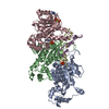
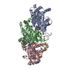
| Title | Yeast Dmc1 post-synaptic complex | ||||||||||||||||||||||||
 Map data Map data | 3.41 angstrom resolution cryoEM structure of yeast Dmc1-dsDNA post-synaptic complex | ||||||||||||||||||||||||
 Sample Sample |
| ||||||||||||||||||||||||
 Keywords Keywords | Meiotic recombination protein DMC1 / DNA BINDING PROTEIN / DNA BINDING PROTEIN-DNA complex | ||||||||||||||||||||||||
| Function / homology | :  Function and homology information Function and homology information | ||||||||||||||||||||||||
| Biological species |  | ||||||||||||||||||||||||
| Method | helical reconstruction / cryo EM / Resolution: 3.41 Å | ||||||||||||||||||||||||
 Authors Authors | Zhao LY / Xu JF / Wang HW | ||||||||||||||||||||||||
| Funding support |  China, 7 items China, 7 items
| ||||||||||||||||||||||||
 Citation Citation |  Journal: Nucleic Acids Res / Year: 2021 Journal: Nucleic Acids Res / Year: 2021Title: Mechanisms of distinctive mismatch tolerance between Rad51 and Dmc1 in homologous recombination. Authors: Jingfei Xu / Lingyun Zhao / Sijia Peng / Huiying Chu / Rui Liang / Meng Tian / Philip P Connell / Guohui Li / Chunlai Chen / Hong-Wei Wang /    Abstract: Homologous recombination (HR) is a primary DNA double-strand breaks (DSBs) repair mechanism. The recombinases Rad51 and Dmc1 are highly conserved in the RecA family; Rad51 is mainly responsible for ...Homologous recombination (HR) is a primary DNA double-strand breaks (DSBs) repair mechanism. The recombinases Rad51 and Dmc1 are highly conserved in the RecA family; Rad51 is mainly responsible for DNA repair in somatic cells during mitosis while Dmc1 only works during meiosis in germ cells. This spatiotemporal difference is probably due to their distinctive mismatch tolerance during HR: Rad51 does not permit HR in the presence of mismatches, whereas Dmc1 can tolerate certain mismatches. Here, the cryo-EM structures of Rad51-DNA and Dmc1-DNA complexes revealed that the major conformational differences between these two proteins are located in their Loop2 regions, which contain invading single-stranded DNA (ssDNA) binding residues and double-stranded DNA (dsDNA) complementary strand binding residues, stabilizing ssDNA and dsDNA in presynaptic and postsynaptic complexes, respectively. By combining molecular dynamic simulation and single-molecule FRET assays, we identified that V273 and D274 in the Loop2 region of human RAD51 (hRAD51), corresponding to P274 and G275 of human DMC1 (hDMC1), are the key residues regulating mismatch tolerance during strand exchange in HR. This HR accuracy control mechanism provides mechanistic insights into the specific roles of Rad51 and Dmc1 in DNA double-strand break repair and may shed light on the regulatory mechanism of genetic recombination in mitosis and meiosis. | ||||||||||||||||||||||||
| History |
|
- Structure visualization
| Supplemental images |
|---|
- Downloads & links
-EMDB archive
| Map data |  emd_31154.map.gz emd_31154.map.gz | 15.2 MB |  EMDB map data format EMDB map data format | |
|---|---|---|---|---|
| Header (meta data) |  emd-31154-v30.xml emd-31154-v30.xml emd-31154.xml emd-31154.xml | 13.5 KB 13.5 KB | Display Display |  EMDB header EMDB header |
| Images |  emd_31154.png emd_31154.png | 214.8 KB | ||
| Filedesc metadata |  emd-31154.cif.gz emd-31154.cif.gz | 5.7 KB | ||
| Archive directory |  http://ftp.pdbj.org/pub/emdb/structures/EMD-31154 http://ftp.pdbj.org/pub/emdb/structures/EMD-31154 ftp://ftp.pdbj.org/pub/emdb/structures/EMD-31154 ftp://ftp.pdbj.org/pub/emdb/structures/EMD-31154 | HTTPS FTP |
-Related structure data
| Related structure data |  7ej7MC  7ej6C  7ejcC  7ejeC M: atomic model generated by this map C: citing same article ( |
|---|---|
| Similar structure data | Similarity search - Function & homology  F&H Search F&H Search |
- Links
Links
| EMDB pages |  EMDB (EBI/PDBe) / EMDB (EBI/PDBe) /  EMDataResource EMDataResource |
|---|
- Map
Map
| File |  Download / File: emd_31154.map.gz / Format: CCP4 / Size: 178 MB / Type: IMAGE STORED AS FLOATING POINT NUMBER (4 BYTES) Download / File: emd_31154.map.gz / Format: CCP4 / Size: 178 MB / Type: IMAGE STORED AS FLOATING POINT NUMBER (4 BYTES) | ||||||||||||||||||||||||||||||||||||
|---|---|---|---|---|---|---|---|---|---|---|---|---|---|---|---|---|---|---|---|---|---|---|---|---|---|---|---|---|---|---|---|---|---|---|---|---|---|
| Annotation | 3.41 angstrom resolution cryoEM structure of yeast Dmc1-dsDNA post-synaptic complex | ||||||||||||||||||||||||||||||||||||
| Projections & slices | Image control
Images are generated by Spider. | ||||||||||||||||||||||||||||||||||||
| Voxel size | X=Y=Z: 1.025 Å | ||||||||||||||||||||||||||||||||||||
| Density |
| ||||||||||||||||||||||||||||||||||||
| Symmetry | Space group: 1 | ||||||||||||||||||||||||||||||||||||
| Details | EMDB XML:
|
-Supplemental data
- Sample components
-Entire : Yeast Dmc1 post-synaptic complex
| Entire | Name: Yeast Dmc1 post-synaptic complex |
|---|---|
| Components |
|
-Supramolecule #1: Yeast Dmc1 post-synaptic complex
| Supramolecule | Name: Yeast Dmc1 post-synaptic complex / type: complex / ID: 1 / Parent: 0 / Macromolecule list: #1-#3 Details: 3.41 angstrom cryoEM structure of yeast Dmc1-dsDNA post-synaptic complex |
|---|---|
| Source (natural) | Organism:  |
-Macromolecule #1: HLJ1_G0016300.mRNA.1.CDS.1
| Macromolecule | Name: HLJ1_G0016300.mRNA.1.CDS.1 / type: protein_or_peptide / ID: 1 / Number of copies: 3 / Enantiomer: LEVO |
|---|---|
| Source (natural) | Organism:  |
| Molecular weight | Theoretical: 36.657539 KDa |
| Recombinant expression | Organism:  |
| Sequence | String: MSVTGTEIDS DTAKNILSVD ELQNYGINAS DLQKLKSGGI YTVNTVLSTT RRHLCKIKGL SEVKVEKIKE AAGKIIQVGF IPATVQLDI RQRVYSLSTG SKQLDSILGG GIMTMSITEV FGEFRCGKTQ MSHTLCVTTQ LPREMGGGEG KVAYIDTEGT F RPERIKQI ...String: MSVTGTEIDS DTAKNILSVD ELQNYGINAS DLQKLKSGGI YTVNTVLSTT RRHLCKIKGL SEVKVEKIKE AAGKIIQVGF IPATVQLDI RQRVYSLSTG SKQLDSILGG GIMTMSITEV FGEFRCGKTQ MSHTLCVTTQ LPREMGGGEG KVAYIDTEGT F RPERIKQI AEGYELDPES CLANVSYARA LNSEHQMELV EQLGEELSSG DYRLIVVDSI MANFRVDYCG RGELSERQQK LN QHLFKLN RLAEEFNVAV FLTNQVQSDP GASALFASAD GRKPIGGHVL AHASATRILL RKGRGDERVA KLQDSPDMPE KEC VYVIGE KGITDSSD UniProtKB: UNIPROTKB: A0A6L0Z498 |
-Macromolecule #2: DNA (5'-D(P*TP*TP*TP*TP*TP*TP*TP*TP*T)-3')
| Macromolecule | Name: DNA (5'-D(P*TP*TP*TP*TP*TP*TP*TP*TP*T)-3') / type: dna / ID: 2 / Number of copies: 1 / Classification: DNA |
|---|---|
| Source (natural) | Organism:  |
| Molecular weight | Theoretical: 2.692778 KDa |
| Sequence | String: (DT)(DT)(DT)(DT)(DT)(DT)(DT)(DT)(DT) |
-Macromolecule #3: DNA (5'-D(P*AP*AP*AP*AP*AP*AP*AP*AP*A)-3')
| Macromolecule | Name: DNA (5'-D(P*AP*AP*AP*AP*AP*AP*AP*AP*A)-3') / type: dna / ID: 3 / Number of copies: 1 / Classification: DNA |
|---|---|
| Source (natural) | Organism:  |
| Molecular weight | Theoretical: 2.773904 KDa |
| Sequence | String: (DA)(DA)(DA)(DA)(DA)(DA)(DA)(DA)(DA) |
-Macromolecule #4: MAGNESIUM ION
| Macromolecule | Name: MAGNESIUM ION / type: ligand / ID: 4 / Number of copies: 3 / Formula: MG |
|---|---|
| Molecular weight | Theoretical: 24.305 Da |
-Macromolecule #5: ADENOSINE-5'-TRIPHOSPHATE
| Macromolecule | Name: ADENOSINE-5'-TRIPHOSPHATE / type: ligand / ID: 5 / Number of copies: 3 / Formula: ATP |
|---|---|
| Molecular weight | Theoretical: 507.181 Da |
| Chemical component information |  ChemComp-ATP: |
-Experimental details
-Structure determination
| Method | cryo EM |
|---|---|
 Processing Processing | helical reconstruction |
| Aggregation state | filament |
- Sample preparation
| Buffer | pH: 7.5 |
|---|---|
| Vitrification | Cryogen name: ETHANE |
- Electron microscopy
| Microscope | FEI TITAN KRIOS |
|---|---|
| Image recording | Film or detector model: GATAN K2 SUMMIT (4k x 4k) / Average electron dose: 48.0 e/Å2 |
| Electron beam | Acceleration voltage: 300 kV / Electron source:  FIELD EMISSION GUN FIELD EMISSION GUN |
| Electron optics | Illumination mode: FLOOD BEAM / Imaging mode: BRIGHT FIELD / Nominal defocus max: 3.5 µm / Nominal defocus min: 1.0 µm |
| Experimental equipment |  Model: Titan Krios / Image courtesy: FEI Company |
- Image processing
| Final reconstruction | Applied symmetry - Helical parameters - Δz: 15.8 Å Applied symmetry - Helical parameters - Δ&Phi: 56.77 ° Applied symmetry - Helical parameters - Axial symmetry: C1 (asymmetric) Resolution.type: BY AUTHOR / Resolution: 3.41 Å / Resolution method: FSC 0.143 CUT-OFF / Number images used: 71192 |
|---|---|
| Startup model | Type of model: EMDB MAP EMDB ID: |
| Final angle assignment | Type: NOT APPLICABLE |
 Movie
Movie Controller


 Z (Sec.)
 Y (Row.)
 X (Col.)


