 Movie
Movie Controller


+ Open data

- Basic information
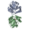
| Entry | Database: EMDB / ID: EMD-31135 | |||||||||
|---|---|---|---|---|---|---|---|---|---|---|
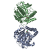
| Title | Bicarbonate transporter complex SbtA-SbtB bound to AMP | |||||||||
 Map data Map data | ||||||||||
 Sample Sample |
| |||||||||
 Keywords Keywords | Bicarbonate transporter / CO2 concentrating mechanism / Allosteric inhibition / Photosynthesis / TRANSPORT PROTEIN | |||||||||
| Function / homology |  Function and homology information Function and homology informationregulation of nitrogen utilization / plasma membrane-derived thylakoid membrane / enzyme regulator activity Similarity search - Function | |||||||||
| Biological species |  | |||||||||
| Method | single particle reconstruction / cryo EM / Resolution: 2.7 Å | |||||||||
 Authors Authors | Fang S / Huang X | |||||||||
 Citation Citation | Journal: Proc Natl Acad Sci U S A / Year: 2021 Title: Molecular mechanism underlying transport and allosteric inhibition of bicarbonate transporter SbtA. Authors: Sunzhenhe Fang / Xiaowei Huang / Xue Zhang / Minhua Zhang / Yahui Hao / Hui Guo / Lu-Ning Liu / Fang Yu / Peng Zhang /   Abstract: SbtA is a high-affinity, sodium-dependent bicarbonate transporter found in the cyanobacterial CO-concentrating mechanism (CCM). SbtA forms a complex with SbtB, while SbtB allosterically regulates the ...SbtA is a high-affinity, sodium-dependent bicarbonate transporter found in the cyanobacterial CO-concentrating mechanism (CCM). SbtA forms a complex with SbtB, while SbtB allosterically regulates the transport activity of SbtA by binding with adenyl nucleotides. The underlying mechanism of transport and regulation of SbtA is largely unknown. In this study, we report the three-dimensional structures of the cyanobacterial sp. PCC 6803 SbtA-SbtB complex in both the presence and absence of HCO and/or AMP at 2.7 Å and 3.2 Å resolution. An analysis of the inward-facing state of the SbtA structure reveals the HCO/Na binding site, providing evidence for the functional unit as a trimer. A structural comparison found that SbtA adopts an elevator mechanism for bicarbonate transport. A structure-based analysis revealed that the allosteric inhibition of SbtA by SbtB occurs mainly through the T-loop of SbtB, which binds to both the core domain and the scaffold domain of SbtA and locks it in an inward-facing state. T-loop conformation is stabilized by the AMP molecules binding at the SbtB trimer interfaces and may be adjusted by other adenyl nucleotides. The unique regulatory mechanism of SbtA by SbtB makes it important to study inorganic carbon uptake systems in CCM, which can be used to modify photosynthesis in crops. | |||||||||
| History |
|
- Structure visualization
| Movie |
Movie viewer |
|---|---|
| Structure viewer | EM map: SurfViewMolmilJmol/JSmol |
| Supplemental images |

- Downloads & links
-EMDB archive
| Map data | emd_31135.map.gz | 4.8 MB | EMDB map data format | |
|---|---|---|---|---|
| Header (meta data) | emd-31135-v30.xmlemd-31135.xml | 15.5 KB 15.5 KB | Display Display | EMDB header |
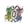
| Images |  emd_31135.png emd_31135.png | 119.8 KB | ||
| Filedesc metadata | emd-31135.cif.gz | 5.4 KB | ||
| Others | emd_31135_half_map_1.map.gzemd_31135_half_map_2.map.gz | 40.6 MB 40.7 MB | ||
| Archive directory |  http://ftp.pdbj.org/pub/emdb/structures/EMD-31135ftp://ftp.pdbj.org/pub/emdb/structures/EMD-31135 http://ftp.pdbj.org/pub/emdb/structures/EMD-31135ftp://ftp.pdbj.org/pub/emdb/structures/EMD-31135 | HTTPS FTP |
-Related structure data
| Related structure data |  7egkMC  7eglC M: atomic model generated by this map C: citing same article ( |
|---|---|
| Similar structure data |


-Links
| EMDB pages | EMDB (EBI/PDBe) / EMDataResource |
|---|
-Map
| File | Download / File: emd_31135.map.gz / Format: CCP4 / Size: 52.7 MB / Type: IMAGE STORED AS FLOATING POINT NUMBER (4 BYTES) | ||||||||||||||||||||||||||||||||||||||||||||||||||||||||||||||||||||
|---|---|---|---|---|---|---|---|---|---|---|---|---|---|---|---|---|---|---|---|---|---|---|---|---|---|---|---|---|---|---|---|---|---|---|---|---|---|---|---|---|---|---|---|---|---|---|---|---|---|---|---|---|---|---|---|---|---|---|---|---|---|---|---|---|---|---|---|---|---|
| Projections & slices | Image control
Images are generated by Spider. | ||||||||||||||||||||||||||||||||||||||||||||||||||||||||||||||||||||
| Voxel size | X=Y=Z: 1.0773 Å | ||||||||||||||||||||||||||||||||||||||||||||||||||||||||||||||||||||
| Density |
| ||||||||||||||||||||||||||||||||||||||||||||||||||||||||||||||||||||
| Symmetry | Space group: 1 | ||||||||||||||||||||||||||||||||||||||||||||||||||||||||||||||||||||
| Details | EMDB XML:
CCP4 map header:
| ||||||||||||||||||||||||||||||||||||||||||||||||||||||||||||||||||||
 Z (Sec.)
 Y (Row.)
 X (Col.)


-Supplemental data
-Half map: #1
| File | emd_31135_half_map_1.map | ||||||||||||
|---|---|---|---|---|---|---|---|---|---|---|---|---|---|
| Projections & Slices |
| ||||||||||||
| Density Histograms |


-Half map: #2
| File | emd_31135_half_map_2.map | ||||||||||||
|---|---|---|---|---|---|---|---|---|---|---|---|---|---|
| Projections & Slices |
| ||||||||||||
| Density Histograms |


- Sample components
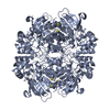
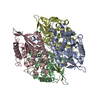
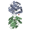
-Entire : Bicarbonate transporter SbtA in complex with regulator SbtB
| Entire | Name: Bicarbonate transporter SbtA in complex with regulator SbtB |
|---|---|
| Components |
|
-Supramolecule #1: Bicarbonate transporter SbtA in complex with regulator SbtB
| Supramolecule | Name: Bicarbonate transporter SbtA in complex with regulator SbtB type: complex / ID: 1 / Parent: 0 / Macromolecule list: #1-#2 |
|---|---|
| Source (natural) | Organism: |
-Macromolecule #1: Sodium-dependent bicarbonate transporter SbtA
| Macromolecule | Name: Sodium-dependent bicarbonate transporter SbtA / type: protein_or_peptide / ID: 1 / Number of copies: 3 / Enantiomer: LEVO |
|---|---|
| Source (natural) | Organism: |
| Molecular weight | Theoretical: 39.671246 KDa |
| Recombinant expression | Organism: |
| Sequence | String: MDFLSNFLTD FVGQLQSPTL AFLIGGMVIA ALGTQLVIPE AISTIIVFML LTKIGLTGGM AIRNSNLTEM LLPVAFSVIL GILIVFIAR FTLAKLPNVR TVDALATGGL FGAVSGSTMA AALTTLEESK ISYEAWAGAL YPFMDIPALV TAIVVANIYL N KRKRKSAA ...String: MDFLSNFLTD FVGQLQSPTL AFLIGGMVIA ALGTQLVIPE AISTIIVFML LTKIGLTGGM AIRNSNLTEM LLPVAFSVIL GILIVFIAR FTLAKLPNVR TVDALATGGL FGAVSGSTMA AALTTLEESK ISYEAWAGAL YPFMDIPALV TAIVVANIYL N KRKRKSAA ASIEESFSKQ PVAAGDYGDQ TDYPRTRQEY LSQQEPEDNR VKIWPIIEES LQGPALSAML LGLALGIFTK PE SVYEGFY DPLFRGLLSI LMLIMGMEAW SRIGELRKVA QWYVVYSLIA PIVHGFIAFG LGMIAHYATG FSLGGVVVLA VIA ASSSDI SGPPTLRAGI PSANPSAYIG SSTAIGTPIA IGVCIPLFIG LAQTLGAG UniProtKB: Slr1512 protein |
-Macromolecule #2: Membrane-associated protein SbtB
| Macromolecule | Name: Membrane-associated protein SbtB / type: protein_or_peptide / ID: 2 / Number of copies: 3 / Enantiomer: LEVO |
|---|---|
| Source (natural) | Organism: |
| Molecular weight | Theoretical: 12.02181 KDa |
| Recombinant expression | Organism: |
| Sequence | String: MAKPANKLVI VTEKILLKKI AKIIDESGAK GYTVMNTGGK GSRNVRSSGQ PNTSDIEANI KFEILTETRE MAEEIADRVA VKYFNDYAG IIYICSAEVL YGHTFCGPEG C UniProtKB: Membrane-associated protein slr1513 |
-Macromolecule #3: SODIUM ION
| Macromolecule | Name: SODIUM ION / type: ligand / ID: 3 / Number of copies: 3 |
|---|---|
| Molecular weight | Theoretical: 22.99 Da |
-Macromolecule #4: ADENOSINE MONOPHOSPHATE
| Macromolecule | Name: ADENOSINE MONOPHOSPHATE / type: ligand / ID: 4 / Number of copies: 3 / Formula: AMP |
|---|---|
| Molecular weight | Theoretical: 347.221 Da |
| Chemical component information |  ChemComp-AMP: |
-Experimental details
-Structure determination
| Method | cryo EM |
|---|---|
 Processing Processing | single particle reconstruction |
| Aggregation state | particle |
-Sample preparation
| Buffer | pH: 8 |
|---|---|
| Grid | Model: Quantifoil R1.2/1.3 / Material: COPPER / Mesh: 300 |
| Vitrification | Cryogen name: ETHANE / Chamber humidity: 100 % / Chamber temperature: 281.15 K / Instrument: FEI VITROBOT MARK IV |
- Electron microscopy
| Microscope | FEI TITAN KRIOS |
|---|---|
| Image recording | Film or detector model: GATAN K3 (6k x 4k) / Average electron dose: 50.0 e/Å2 |
| Electron beam | Acceleration voltage: 300 kV / Electron source:  FIELD EMISSION GUN FIELD EMISSION GUN |
| Electron optics | Illumination mode: SPOT SCAN / Imaging mode: BRIGHT FIELD |
| Experimental equipment |  Model: Titan Krios / Image courtesy: FEI Company |
-Image processing
| Startup model | Type of model: NONE |
|---|---|
| Final reconstruction | Applied symmetry - Point group: C3 (3 fold cyclic) / Resolution.type: BY AUTHOR / Resolution: 2.7 Å / Resolution method: FSC 0.143 CUT-OFF / Number images used: 125998 |
| Initial angle assignment | Type: MAXIMUM LIKELIHOOD |
| Final angle assignment | Type: MAXIMUM LIKELIHOOD |
