 Movie
Movie Controller


+ Open data

- Basic information
| Entry | Database: EMDB / ID: EMD-30646 | |||||||||
|---|---|---|---|---|---|---|---|---|---|---|
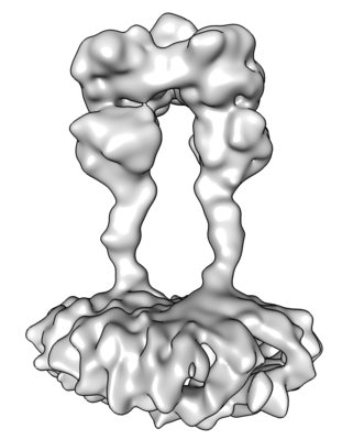
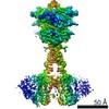
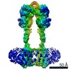
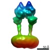
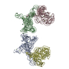
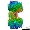
| Title | Structure of Calcium-Sensing Receptor in an inactive state | |||||||||
 Map data Map data | ||||||||||
 Sample Sample |
| |||||||||
| Biological species |  | |||||||||
| Method | single particle reconstruction / cryo EM / Resolution: 7.2 Å | |||||||||
 Authors Authors | Wen TL / Yang X / Shen YQ | |||||||||
 Citation Citation | Journal: Sci Adv / Year: 2021 Title: Structural basis for activation and allosteric modulation of full-length calcium-sensing receptor. Authors: Tianlei Wen / Ziyu Wang / Xiaozhe Chen / Yue Ren / Xuhang Lu / Yangfei Xing / Jing Lu / Shenghai Chang / Xing Zhang / Yuequan Shen / Xue Yang /  Abstract: Calcium-sensing receptor (CaSR) is a class C G protein-coupled receptor (GPCR) that plays an important role in calcium homeostasis and parathyroid hormone secretion. Here, we present multiple cryo- ...Calcium-sensing receptor (CaSR) is a class C G protein-coupled receptor (GPCR) that plays an important role in calcium homeostasis and parathyroid hormone secretion. Here, we present multiple cryo-electron microscopy structures of full-length CaSR in distinct ligand-bound states. Ligands (Ca and l-tryptophan) bind to the extracellular domain of CaSR and induce large-scale conformational changes, leading to the closure of two heptahelical transmembrane domains (7TMDs) for activation. The positive modulator (evocalcet) and the negative allosteric modulator (NPS-2143) occupy the similar binding pocket in 7TMD. The binding of NPS-2143 causes a considerable rearrangement of two 7TMDs, forming an inactivated TM6/TM6 interface. Moreover, a total of 305 disease-causing missense mutations of CaSR have been mapped to the structure in the active state, creating hotspot maps of five clinical endocrine disorders. Our results provide a structural framework for understanding the activation, allosteric modulation mechanism, and disease therapy for class C GPCRs. | |||||||||
| History |
|
- Structure visualization
| Movie |
 Movie viewer Movie viewer |
|---|---|
| Structure viewer | EM map: SurfViewMolmilJmol/JSmol |
| Supplemental images |
- Downloads & links
-EMDB archive
| Map data | emd_30646.map.gz | 60.2 MB | EMDB map data format | |
|---|---|---|---|---|
| Header (meta data) | emd-30646-v30.xmlemd-30646.xml | 10 KB 10 KB | Display Display | EMDB header |
| Images |  emd_30646.png emd_30646.png | 48.5 KB | ||
| Archive directory |  http://ftp.pdbj.org/pub/emdb/structures/EMD-30646ftp://ftp.pdbj.org/pub/emdb/structures/EMD-30646 http://ftp.pdbj.org/pub/emdb/structures/EMD-30646ftp://ftp.pdbj.org/pub/emdb/structures/EMD-30646 | HTTPS FTP |
-Related structure data


-Links
| EMDB pages | EMDB (EBI/PDBe) / EMDataResource |
|---|
-Map
| File | Download / File: emd_30646.map.gz / Format: CCP4 / Size: 125 MB / Type: IMAGE STORED AS FLOATING POINT NUMBER (4 BYTES) | ||||||||||||||||||||||||||||||||||||||||||||||||||||||||||||||||||||
|---|---|---|---|---|---|---|---|---|---|---|---|---|---|---|---|---|---|---|---|---|---|---|---|---|---|---|---|---|---|---|---|---|---|---|---|---|---|---|---|---|---|---|---|---|---|---|---|---|---|---|---|---|---|---|---|---|---|---|---|---|---|---|---|---|---|---|---|---|---|
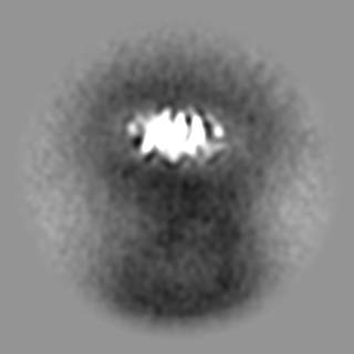
| Projections & slices | Image control
Images are generated by Spider. | ||||||||||||||||||||||||||||||||||||||||||||||||||||||||||||||||||||
| Voxel size | X=Y=Z: 1.014 Å | ||||||||||||||||||||||||||||||||||||||||||||||||||||||||||||||||||||
| Density |
| ||||||||||||||||||||||||||||||||||||||||||||||||||||||||||||||||||||
| Symmetry | Space group: 1 | ||||||||||||||||||||||||||||||||||||||||||||||||||||||||||||||||||||
| Details | EMDB XML:
CCP4 map header:
| ||||||||||||||||||||||||||||||||||||||||||||||||||||||||||||||||||||
 Z (Sec.)
 Y (Row.)
 X (Col.)


-Supplemental data
- Sample components
-Entire : Calcium-Sensing Receptor in an inactive state
| Entire | Name: Calcium-Sensing Receptor in an inactive state |
|---|---|
| Components |
|
-Supramolecule #1: Calcium-Sensing Receptor in an inactive state
| Supramolecule | Name: Calcium-Sensing Receptor in an inactive state / type: cell / ID: 1 / Parent: 0 / Macromolecule list: all |
|---|---|
| Source (natural) | Organism: |
-Macromolecule #1: Calcium-Sensing Receptor, CaSR
| Macromolecule | Name: Calcium-Sensing Receptor, CaSR / type: protein_or_peptide / ID: 1 / Enantiomer: LEVO |
|---|---|
| Source (natural) | Organism: |
| Recombinant expression | Organism:  Homo sapiens (human) Homo sapiens (human) |
| Sequence | String: MTLYSCCLIL LLFTWNTAAY GPNQRAQKKG DIILGGLFPI HFGVAAKDQD LKSRPESVEC IRYNFRGFRW LQAMIFAIEE INNSPNLLPN MTLGYRIFDT CNTVSKALEA TLSFVAQNKI DSLNLDEFCN CSEHIPSTIA VVGATGSGVS TAVANLLGLF YIPQVSYASS ...String: MTLYSCCLIL LLFTWNTAAY GPNQRAQKKG DIILGGLFPI HFGVAAKDQD LKSRPESVEC IRYNFRGFRW LQAMIFAIEE INNSPNLLPN MTLGYRIFDT CNTVSKALEA TLSFVAQNKI DSLNLDEFCN CSEHIPSTIA VVGATGSGVS TAVANLLGLF YIPQVSYASS SRLLSNKNQF KSFLRTIPND EHQATAMADI IEYFRWNWVG TIAADDDYGR PGIEKFREEA EERDICIDFS ELISQYSDEE EIQQVVEVIQ NSTARVIVVF SSGPDLEPLI KEIVRRNITG KIWLASEAWA SSSLIAMPEF FRVIGSTIGF ALKAGQIPGF REFLQKVHPK KSANNGFAKE FWEETFNCYL PSESKNSPAS ASFHKAHEEG LGAGNGTAAF RPPCTGDENI TSVETPYMDF THLRISYNVY LAVYSIAHAL QDIYTCTPGK GLFTNGSCAD IKKVEAWQVL KHLRHLNFTS NMGEQVDFDE FGDLVGNYSI INWHLSPEDG SVVFEEVGHY NVYAKKGERL FINENKILWS GFSKEVPFSN CSRDCLPGTR KGIIEGEPTC CFECVDCPDG EYSDETDASA CDKCPEDYWS NENHTSCIPK QIEFLSWTEP FGIALTLFAV LGIFLTSFVL GVFTKFRNTP IVKATNRELS YLLLFSLLCC FSSSLFFIGE PQNWTCRLRQ PAFGISFVLC ISCILVKTNR VLLVFEAKIP TSLHRKWWGL NLQFLLVFLC TFVQIVICVI WLYTAPPSSY RNHELEDEII FITCHEGSLM ALGFLIGYTC LLAAICFFFA FKSRKLPENF NEAKFITFSM LIFFIVWISF IPAYASTYGK FVSAVEVIAI LAASFGLLAC IFFNKVYIIL FKPSRNTIEE VRCSTAAHAF KVAARATLRR SNVSRKRSNS LGGSTGSTPS SSISSKSNHE DPFPLPASAE RQRQQQRGCK QKVSFGSGTV TLSLSFEEPQ KNAMANRNAK RRNSLEAQNS DDSLMRHRAL LALQNSESLS AEPGFQTASS PETSSQESVV GDNKEEVPNP EAEPSLPSAN SRNFIGTGGS SVTENTVHSL ESALEVLFQ |
-Experimental details
-Structure determination
| Method | cryo EM |
|---|---|
 Processing Processing | single particle reconstruction |
| Aggregation state | particle |
-Sample preparation
| Buffer | pH: 7.5 |
|---|---|
| Grid | Model: Quantifoil R2/1 / Material: COPPER / Mesh: 200 / Pretreatment - Type: GLOW DISCHARGE |
| Vitrification | Cryogen name: ETHANE / Chamber humidity: 100 % |
- Electron microscopy
| Microscope | FEI TITAN |
|---|---|
| Image recording | Film or detector model: GATAN K2 SUMMIT (4k x 4k) / Detector mode: COUNTING / Average electron dose: 50.0 e/Å2 |
| Electron beam | Acceleration voltage: 300 kV / Electron source:  FIELD EMISSION GUN FIELD EMISSION GUN |
| Electron optics | Illumination mode: FLOOD BEAM / Imaging mode: BRIGHT FIELD / Nominal defocus max: 25.0 µm / Nominal defocus min: 5.0 µm |
| Sample stage | Specimen holder model: FEI TITAN KRIOS AUTOGRID HOLDER / Cooling holder cryogen: NITROGEN |
-Image processing
| Final reconstruction | Resolution.type: BY AUTHOR / Resolution: 7.2 Å / Resolution method: FSC 0.143 CUT-OFF / Number images used: 80104 |
|---|---|
| Initial angle assignment | Type: NOT APPLICABLE |
| Final angle assignment | Type: NOT APPLICABLE |
-Atomic model buiding 1
| Refinement | Space: REAL / Protocol: AB INITIO MODEL |
|---|
