 Movie
Movie Controller

[English] 日本語
 Yorodumi
- PDB-1x8w: Structure of the Tetrahymena Ribozyme: Base Triple Sandwich and M... -
+ Open data

- Basic information
| Entry | Database: PDB / ID: 1x8w | ||||||
|---|---|---|---|---|---|---|---|
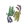
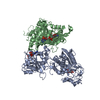
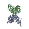
| Title | Structure of the Tetrahymena Ribozyme: Base Triple Sandwich and Metal Ion at the Active Site | ||||||
 Components Components | Tetrahymena ribozyme RNA | ||||||
 Keywords Keywords | RNA / Catalytic RNA / ribozyme / group I intron / guanosine binding site / metal ions / active site / catalytic mechanism / base triples / conformational changes | ||||||
| Function / homology | RNA / RNA (> 10) / RNA (> 100) Function and homology information Function and homology information | ||||||
| Method |  X-RAY DIFFRACTION / SYNCHROTRON / MIR / Resolution: 3.8 Å X-RAY DIFFRACTION / SYNCHROTRON / MIR / Resolution: 3.8 Å | ||||||
 Authors Authors | Guo, F. / Gooding, A.R. / Cech, T.R. | ||||||
 Citation Citation | Journal: Mol.Cell / Year: 2004 Title: Structure of the Tetrahymena ribozyme: base triple sandwich and metal ion at the active site. Authors: Guo, F. / Gooding, A.R. / Cech, T.R. | ||||||
| History |
|
- Structure visualization
| Structure viewer | Molecule: MolmilJmol/JSmol |
|---|
- Downloads & links
-Download
| PDBx/mmCIF format | 1x8w.cif.gz | 464 KB | Display | PDBx/mmCIF format |
|---|---|---|---|---|
| PDB format | pdb1x8w.ent.gz | 358.1 KB | Display | PDB format |
| PDBx/mmJSON format | 1x8w.json.gz | Tree view | PDBx/mmJSON format | |
| Others |  Other downloads Other downloads |
-Validation report
| Arichive directory | https://data.pdbj.org/pub/pdb/validation_reports/x8/1x8wftp://data.pdbj.org/pub/pdb/validation_reports/x8/1x8w | HTTPS FTP |
|---|
-Related structure data
| Similar structure data |
|---|


-Links
 PDBj
PDBj


- Assembly
| Deposited unit | 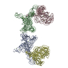
| ||||||||
|---|---|---|---|---|---|---|---|---|---|
| 1 | 
| ||||||||
| 2 | 
| ||||||||
| 3 | 
| ||||||||
| 4 | 
| ||||||||
| Unit cell |
|
-Components
| #1: RNA chain | Mass: 79966.438 Da / Num. of mol.: 4 / Mutation: A210G, U259A, A269G, U277C, A304G / Source method: obtained synthetically Details: RNA WAS PREPARED BY IN VITRO TRANSCRIPTION. The sequence of this RNA can be found naturally in TETRAHYMENA THERMOPHILA. #2: Chemical | ChemComp-MG /   Mass: 24.305 Da / Num. of mol.: 38 / Source method: obtained synthetically / Formula: Mg Mass: 24.305 Da / Num. of mol.: 38 / Source method: obtained synthetically / Formula: Mg |
|---|
-Experimental details
-Experiment
| Experiment | Method: X-RAY DIFFRACTION / Number of used crystals: 3 |
|---|
- Sample preparation
| Crystal | Density Matthews: 3.6 Å3/Da / Density % sol: 70 % | ||||||||||||||||||||||||||||||||||||||||||||||||||||
|---|---|---|---|---|---|---|---|---|---|---|---|---|---|---|---|---|---|---|---|---|---|---|---|---|---|---|---|---|---|---|---|---|---|---|---|---|---|---|---|---|---|---|---|---|---|---|---|---|---|---|---|---|---|
| Crystal grow | Temperature: 303 K / Method: vapor diffusion, hanging drop / pH: 6 Details: 9.5% 2-methyl-2,4-pentanediol (MPD), 25 mM K cacodylate pH 6.0, 25 mM KCl, 5mM NaCl, 17.5 mM MgCl2 and 0.25 mM spermine, VAPOR DIFFUSION, HANGING DROP, temperature 303K | ||||||||||||||||||||||||||||||||||||||||||||||||||||
| Components of the solutions |
|
-Data collection
| Diffraction | Mean temperature: 100 K |
|---|---|
| Diffraction source | Source: SYNCHROTRON / Site: ALS  / Beamline: 8.2.2 / Wavelength: 1.0332 Å / Beamline: 8.2.2 / Wavelength: 1.0332 Å |
| Detector | Type: ADSC QUANTUM 315 / Detector: CCD / Date: Oct 30, 2002 / Details: Double crystal, Si(111) |
| Radiation | Monochromator: Si 111 Channel / Protocol: SINGLE WAVELENGTH / Monochromatic (M) / Laue (L): M / Scattering type: x-ray |
| Radiation wavelength | Wavelength: 1.0332 Å / Relative weight: 1 |
| Reflection | Resolution: 3.8→20 Å / Num. all: 43985 / Num. obs: 43972 / % possible obs: 93.9 % / Observed criterion σ(F): 0.01 / Observed criterion σ(I): -3 / Redundancy: 4.8 % / Rmerge(I) obs: 0.054 / Net I/σ(I): 24 |
| Reflection shell | Resolution: 3.8→3.97 Å / Redundancy: 1.8 % / Rmerge(I) obs: 0.255 / Mean I/σ(I) obs: 3.6 / Num. unique all: 4429 / % possible all: 76.8 |
- Processing
| Software |
| ||||||||||||||||||||
|---|---|---|---|---|---|---|---|---|---|---|---|---|---|---|---|---|---|---|---|---|---|
| Refinement | Method to determine structure: MIR / Resolution: 3.8→20 Å / Isotropic thermal model: Isotropic / Cross valid method: THROUGHOUT / σ(F): 0 / Stereochemistry target values: Engh & Huber Details: experimental phase probability distribution is used in the maximum likehood target
| ||||||||||||||||||||
| Displacement parameters | Biso mean: 122 Å2 | ||||||||||||||||||||
| Refine analyze |
| ||||||||||||||||||||
| Refinement step | Cycle: LAST / Resolution: 3.8→20 Å
| ||||||||||||||||||||
| Refine LS restraints |
| ||||||||||||||||||||
| LS refinement shell | Resolution: 3.8→3.97 Å / Rfactor Rfree error: 0.022
|
