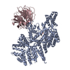
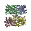
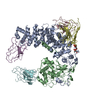
Glycoside hydrolase, family 14B, plant / Beta-amylase active site 2. / Glycoside hydrolase, family 14, conserved site / Beta-amylase active site 1. / Glycoside hydrolase, family 14 / Glycosyl hydrolase family 14 / Glycoside hydrolase superfamily Similarity search - Domain/homology
Journal: Structure / Year: 2021 Title: Improving particle quality in cryo-EM analysis using a PEGylation method. Authors: Zhikuan Zhang / Hideki Shigematsu / Toshiyuki Shimizu / Umeharu Ohto / Abstract: Cryo-electron microscopy (cryo-EM) is widely used for structural biology studies and has been developed extensively in recent years. However, its sample vitrification process is a major limitation ...Cryo-electron microscopy (cryo-EM) is widely used for structural biology studies and has been developed extensively in recent years. However, its sample vitrification process is a major limitation because it causes severe particle aggregation and/or denaturation. This effect is thought to occur because particles tend to stick to the "deadly" air-water interface during vitrification. Here, we report a method for PEGylation of proteins that can efficiently protect particles against such problems during vitrification. This method alleviates the laborious process of fine-tuning the vitrification conditions, allowing for analysis of samples that would otherwise be discarded.
History
Deposition
Jul 26, 2020
-
Header (metadata) release
Jun 16, 2021
-
Map release
Jun 16, 2021
-
Update
Oct 20, 2021
-
Current status
Oct 20, 2021
Processing site: PDBj / Status: Released
-
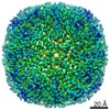
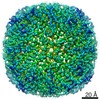
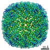
Structure visualization
Movie
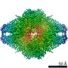
Surface view with section colored by density value
In the structure databanks used in Yorodumi, some data are registered as the other names, "COVID-19 virus" and "2019-nCoV". Here are the details of the virus and the list of structure data.
Jan 31, 2019. EMDB accession codes are about to change! (news from PDBe EMDB page)
EMDB accession codes are about to change! (news from PDBe EMDB page)
The allocation of 4 digits for EMDB accession codes will soon come to an end. Whilst these codes will remain in use, new EMDB accession codes will include an additional digit and will expand incrementally as the available range of codes is exhausted. The current 4-digit format prefixed with “EMD-” (i.e. EMD-XXXX) will advance to a 5-digit format (i.e. EMD-XXXXX), and so on. It is currently estimated that the 4-digit codes will be depleted around Spring 2019, at which point the 5-digit format will come into force.
The EM Navigator/Yorodumi systems omit the EMD- prefix.
Related info.:Q: What is EMD? / ID/Accession-code notation in Yorodumi/EM Navigator
Yorodumi is a browser for structure data from EMDB, PDB, SASBDB, etc.
This page is also the successor to EM Navigator detail page, and also detail information page/front-end page for Omokage search.
The word "yorodu" (or yorozu) is an old Japanese word meaning "ten thousand". "mi" (miru) is to see.
Related info.:EMDB / PDB / SASBDB / Comparison of 3 databanks / Yorodumi Search / Aug 31, 2016. New EM Navigator & Yorodumi / Yorodumi Papers / Jmol/JSmol / Function and homology information / Changes in new EM Navigator and Yorodumi
 Movie
Movie Controller


 Open data
 Basic information
 Map data
 Sample
 Function and homology information
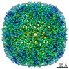
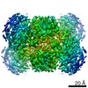
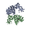
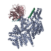
 Ipomoea batatas (sweet potato)
 Authors
 Citation
 Structure visualization
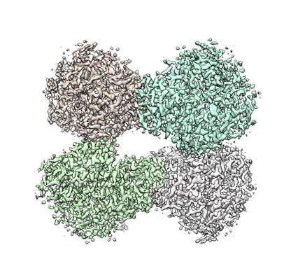
 Downloads & links
 emd_30405.png
 http://ftp.pdbj.org/pub/emdb/structures/EMD-30405


 Z (Sec.)
 Y (Row.)
 X (Col.)


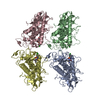
 Sample components
 Spodoptera frugiperda (fall armyworm)
 Processing
 Electron microscopy
 FIELD EMISSION GUN
