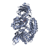
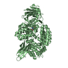
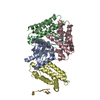
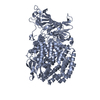
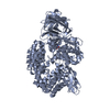
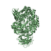
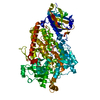
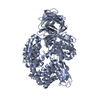
Journal: Cell Discov / Year: 2020 Title: Broad host range of SARS-CoV-2 and the molecular basis for SARS-CoV-2 binding to cat ACE2. Authors: Lili Wu / Qian Chen / Kefang Liu / Jia Wang / Pengcheng Han / Yanfang Zhang / Yu Hu / Yumin Meng / Xiaoqian Pan / Chengpeng Qiao / Siyu Tian / Pei Du / Hao Song / Weifeng Shi / Jianxun Qi / ...Authors: Lili Wu / Qian Chen / Kefang Liu / Jia Wang / Pengcheng Han / Yanfang Zhang / Yu Hu / Yumin Meng / Xiaoqian Pan / Chengpeng Qiao / Siyu Tian / Pei Du / Hao Song / Weifeng Shi / Jianxun Qi / Hong-Wei Wang / Jinghua Yan / George Fu Gao / Qihui Wang / Abstract: Severe acute respiratory syndrome coronavirus 2 (SARS-CoV-2), the causative agent of the recent pandemic COVID-19, is reported to have originated from bats, with its intermediate host unknown to date. ...Severe acute respiratory syndrome coronavirus 2 (SARS-CoV-2), the causative agent of the recent pandemic COVID-19, is reported to have originated from bats, with its intermediate host unknown to date. Here, we screened 26 animal counterparts of the human ACE2 (hACE2), the receptor for SARS-CoV-2 and SARS-CoV, and found that the ACE2s from various species, including pets, domestic animals and multiple wild animals, could bind to SARS-CoV-2 receptor binding domain (RBD) and facilitate the transduction of SARS-CoV-2 pseudovirus. Comparing to SARS-CoV-2, SARS-CoV seems to have a slightly wider range in choosing its receptor. We further resolved the cryo-electron microscopy (cryo-EM) structure of the cat ACE2 (cACE2) in complex with the SARS-CoV-2 RBD at a resolution of 3 Å, revealing similar binding mode as hACE2 to the SARS-CoV-2 RBD. These results shed light on pursuing the intermediate host of SARS-CoV-2 and highlight the necessity of monitoring susceptible hosts to prevent further outbreaks.
History
Deposition
May 29, 2020
-
Header (metadata) release
Sep 2, 2020
-
Map release
Sep 2, 2020
-
Update
Jun 25, 2025
-
Current status
Jun 25, 2025
Processing site: PDBj / Status: Released
-
Structure visualization
Movie
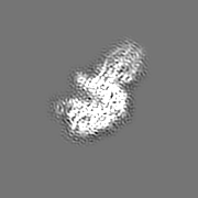
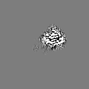
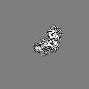
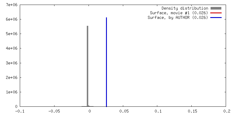
Surface view with section colored by density value
In the structure databanks used in Yorodumi, some data are registered as the other names, "COVID-19 virus" and "2019-nCoV". Here are the details of the virus and the list of structure data.
Jan 31, 2019. EMDB accession codes are about to change! (news from PDBe EMDB page)
EMDB accession codes are about to change! (news from PDBe EMDB page)
The allocation of 4 digits for EMDB accession codes will soon come to an end. Whilst these codes will remain in use, new EMDB accession codes will include an additional digit and will expand incrementally as the available range of codes is exhausted. The current 4-digit format prefixed with “EMD-” (i.e. EMD-XXXX) will advance to a 5-digit format (i.e. EMD-XXXXX), and so on. It is currently estimated that the 4-digit codes will be depleted around Spring 2019, at which point the 5-digit format will come into force.
The EM Navigator/Yorodumi systems omit the EMD- prefix.
Related info.:Q: What is EMD? / ID/Accession-code notation in Yorodumi/EM Navigator
Yorodumi is a browser for structure data from EMDB, PDB, SASBDB, etc.
This page is also the successor to EM Navigator detail page, and also detail information page/front-end page for Omokage search.
The word "yorodu" (or yorozu) is an old Japanese word meaning "ten thousand". "mi" (miru) is to see.
Related info.:EMDB / PDB / SASBDB / Comparison of 3 databanks / Yorodumi Search / Aug 31, 2016. New EM Navigator & Yorodumi / Yorodumi Papers / Jmol/JSmol / Function and homology information / Changes in new EM Navigator and Yorodumi
 Movie
Movie Controller


 Open data
 Basic information
 Map data
 Sample
 Keywords
 Function and homology information

 Severe acute respiratory syndrome coronavirus 2
 Authors
 China, 1 items
  Citation
 Structure visualization
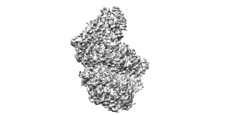
 Downloads & links
 emd_30305.png
 http://ftp.pdbj.org/pub/emdb/structures/EMD-30305


 Z (Sec.)
 Y (Row.)
 X (Col.)


 Sample components

 Spodoptera frugiperda (fall armyworm)
 Processing
 Electron microscopy
 FIELD EMISSION GUN

