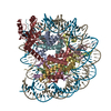
Journal: J Mol Biol / Year: 2021 Title: A Novel N-terminal Region to Chromodomain in CHD7 is Required for the Efficient Remodeling Activity. Authors: Eunhye Lee / Chanshin Kang / Pasi Purhonen / Hans Hebert / Karim Bouazoune / Sungchul Hohng / Ji-Joon Song / Abstract: Chromodomain-Helicase DNA binding protein 7 (CHD7) is an ATP dependent chromatin remodeler involved in maintaining open chromatin structure. Mutations of CHD7 gene causes multiple developmental ...Chromodomain-Helicase DNA binding protein 7 (CHD7) is an ATP dependent chromatin remodeler involved in maintaining open chromatin structure. Mutations of CHD7 gene causes multiple developmental disorders, notably CHARGE syndrome. However, there is not much known about the molecular mechanism by which CHD7 remodels nucleosomes. Here, we performed biochemical and biophysical analysis on CHD7 chromatin remodeler and uncover that N-terminal to the Chromodomain (N-CRD) interacts with nucleosome and contains a high conserved arginine stretch, which is reminiscent of arginine anchor. Importantly, this region is required for efficient ATPase stimulation and nucleosome remodeling activity of CHD7. Furthermore, smFRET analysis shows the mutations in the N-CRD causes the defects in remodeling activity. Collectively, our results uncover the functional importance of a previously unidentified N-terminal region in CHD7 and implicate that the multiple domains in chromatin remodelers are involved in regulating their activities.
History
Deposition
May 25, 2020
-
Header (metadata) release
Jul 28, 2021
-
Map release
Jul 28, 2021
-
Update
Aug 10, 2022
-
Current status
Aug 10, 2022
Processing site: PDBj / Status: Released
-
Structure visualization
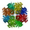
Movie
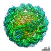
Surface view with section colored by density value
In the structure databanks used in Yorodumi, some data are registered as the other names, "COVID-19 virus" and "2019-nCoV". Here are the details of the virus and the list of structure data.
Jan 31, 2019. EMDB accession codes are about to change! (news from PDBe EMDB page)
EMDB accession codes are about to change! (news from PDBe EMDB page)
The allocation of 4 digits for EMDB accession codes will soon come to an end. Whilst these codes will remain in use, new EMDB accession codes will include an additional digit and will expand incrementally as the available range of codes is exhausted. The current 4-digit format prefixed with “EMD-” (i.e. EMD-XXXX) will advance to a 5-digit format (i.e. EMD-XXXXX), and so on. It is currently estimated that the 4-digit codes will be depleted around Spring 2019, at which point the 5-digit format will come into force.
The EM Navigator/Yorodumi systems omit the EMD- prefix.
Related info.:Q: What is EMD? / ID/Accession-code notation in Yorodumi/EM Navigator
Yorodumi is a browser for structure data from EMDB, PDB, SASBDB, etc.
This page is also the successor to EM Navigator detail page, and also detail information page/front-end page for Omokage search.
The word "yorodu" (or yorozu) is an old Japanese word meaning "ten thousand". "mi" (miru) is to see.
Related info.:EMDB / PDB / SASBDB / Comparison of 3 databanks / Yorodumi Search / Aug 31, 2016. New EM Navigator & Yorodumi / Yorodumi Papers / Jmol/JSmol / Function and homology information / Changes in new EM Navigator and Yorodumi
 Movie
Movie Controller


 Open data
 Basic information
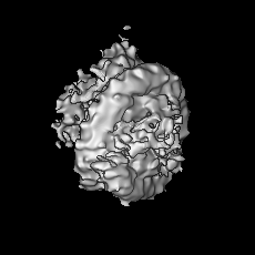
 Map data
 Sample
 Homo sapiens (human) /
  Authors
 Korea, Republic Of, 2 items
  Citation

 Structure visualization
 Movie viewer
 Downloads & links
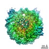
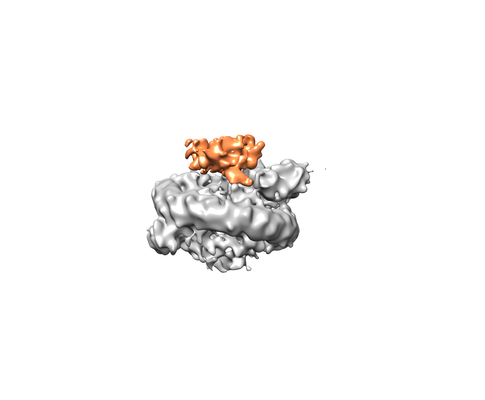 emd_30298.png
 http://ftp.pdbj.org/pub/emdb/structures/EMD-30298
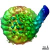


 Z (Sec.)
 Y (Row.)
 X (Col.)


 Sample components
 Processing
 Electron microscopy
 FIELD EMISSION GUN