 ムービー
ムービー コントローラー


+ データを開く

- 基本情報
基本情報
| 登録情報 | データベース: EMDB / ID: EMD-3028 | |||||||||
|---|---|---|---|---|---|---|---|---|---|---|
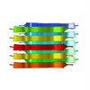
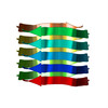
| タイトル | MicroED structure of the toxic core segment, GAVVTGVTAVA, from Parkinson's disease protein, alpha-synuclein, residues 69-78. | |||||||||
 マップデータ マップデータ | microED map of toxic NACore segment, residues 68-78 of alpha-synuclein | |||||||||
 試料 試料 |
| |||||||||
 キーワード キーワード | Amyloid fibrils / alpha-synuclein / MicroED Crystallography / Parkinson's Disease / Peptide / Toxicity | |||||||||
| 機能・相同性 |  機能・相同性情報 機能・相同性情報negative regulation of mitochondrial electron transport, NADH to ubiquinone / : / neutral lipid metabolic process / regulation of acyl-CoA biosynthetic process / negative regulation of dopamine uptake involved in synaptic transmission / negative regulation of norepinephrine uptake / response to desipramine / positive regulation of SNARE complex assembly / positive regulation of hydrogen peroxide catabolic process / supramolecular fiber ...negative regulation of mitochondrial electron transport, NADH to ubiquinone / : / neutral lipid metabolic process / regulation of acyl-CoA biosynthetic process / negative regulation of dopamine uptake involved in synaptic transmission / negative regulation of norepinephrine uptake / response to desipramine / positive regulation of SNARE complex assembly / positive regulation of hydrogen peroxide catabolic process / supramolecular fiber / regulation of synaptic vesicle recycling / mitochondrial membrane organization / negative regulation of chaperone-mediated autophagy / regulation of reactive oxygen species biosynthetic process / negative regulation of platelet-derived growth factor receptor signaling pathway / positive regulation of protein localization to cell periphery / negative regulation of exocytosis / regulation of glutamate secretion / dopamine biosynthetic process / response to iron(II) ion / SNARE complex assembly / positive regulation of neurotransmitter secretion / regulation of locomotion / negative regulation of dopamine metabolic process / positive regulation of inositol phosphate biosynthetic process / regulation of macrophage activation / negative regulation of microtubule polymerization / regulation of norepinephrine uptake / synaptic vesicle transport / transporter regulator activity / synaptic vesicle priming / dopamine uptake involved in synaptic transmission / protein kinase inhibitor activity / mitochondrial ATP synthesis coupled electron transport / regulation of dopamine secretion / dynein complex binding / negative regulation of thrombin-activated receptor signaling pathway / positive regulation of receptor recycling / cuprous ion binding / nuclear outer membrane / response to magnesium ion / positive regulation of endocytosis / positive regulation of exocytosis / synaptic vesicle exocytosis / kinesin binding / synaptic vesicle endocytosis / enzyme inhibitor activity / cysteine-type endopeptidase inhibitor activity / negative regulation of serotonin uptake / response to type II interferon / regulation of presynapse assembly / alpha-tubulin binding / beta-tubulin binding / phospholipase binding / behavioral response to cocaine / supramolecular fiber organization / phospholipid metabolic process / cellular response to fibroblast growth factor stimulus / inclusion body / axon terminus / Hsp70 protein binding / cellular response to epinephrine stimulus / response to interleukin-1 / regulation of microtubule cytoskeleton organization / cellular response to copper ion / positive regulation of release of sequestered calcium ion into cytosol / adult locomotory behavior / SNARE binding / excitatory postsynaptic potential / protein tetramerization / phosphoprotein binding / microglial cell activation / fatty acid metabolic process / ferrous iron binding / regulation of long-term neuronal synaptic plasticity / synapse organization / protein destabilization / PKR-mediated signaling / phospholipid binding / receptor internalization / tau protein binding / long-term synaptic potentiation / terminal bouton / positive regulation of inflammatory response / synaptic vesicle membrane / actin cytoskeleton / actin binding / growth cone / cellular response to oxidative stress / neuron apoptotic process / cell cortex / histone binding / response to lipopolysaccharide / microtubule binding / molecular adaptor activity / chemical synaptic transmission / amyloid fibril formation / negative regulation of neuron apoptotic process / mitochondrial outer membrane / oxidoreductase activity 類似検索 - 分子機能 | |||||||||
| 生物種 |  Homo sapiens (ヒト) Homo sapiens (ヒト) | |||||||||
| 手法 | 電子線結晶学 / クライオ電子顕微鏡法 / 解像度: 1.4 Å | |||||||||
 データ登録者 データ登録者 | Rodriguez JA / Ivanova M / Sawaya MR / Cascio D / Reyes F / Shi D / Johnson L / Guenther E / Zhang M / Jiang L ...Rodriguez JA / Ivanova M / Sawaya MR / Cascio D / Reyes F / Shi D / Johnson L / Guenther E / Zhang M / Jiang L / Arbing MA / Sangwan S / Hattne J / Whitelegge J / Brewster A / Messerschmidt M / Boutet S / Sauter NK / Nannenga B / Gonen T / Eisenberg D | |||||||||
 引用 引用 | ジャーナル: Nature / 年: 2015 タイトル: Structure of the toxic core of α-synuclein from invisible crystals. 著者: Jose A Rodriguez / Magdalena I Ivanova / Michael R Sawaya / Duilio Cascio / Francis E Reyes / Dan Shi / Smriti Sangwan / Elizabeth L Guenther / Lisa M Johnson / Meng Zhang / Lin Jiang / Mark ...著者: Jose A Rodriguez / Magdalena I Ivanova / Michael R Sawaya / Duilio Cascio / Francis E Reyes / Dan Shi / Smriti Sangwan / Elizabeth L Guenther / Lisa M Johnson / Meng Zhang / Lin Jiang / Mark A Arbing / Brent L Nannenga / Johan Hattne / Julian Whitelegge / Aaron S Brewster / Marc Messerschmidt / Sébastien Boutet / Nicholas K Sauter / Tamir Gonen / David S Eisenberg /  要旨: The protein α-synuclein is the main component of Lewy bodies, the neuron-associated aggregates seen in Parkinson disease and other neurodegenerative pathologies. An 11-residue segment, which we term ...The protein α-synuclein is the main component of Lewy bodies, the neuron-associated aggregates seen in Parkinson disease and other neurodegenerative pathologies. An 11-residue segment, which we term NACore, appears to be responsible for amyloid formation and cytotoxicity of human α-synuclein. Here we describe crystals of NACore that have dimensions smaller than the wavelength of visible light and thus are invisible by optical microscopy. As the crystals are thousands of times too small for structure determination by synchrotron X-ray diffraction, we use micro-electron diffraction to determine the structure at atomic resolution. The 1.4 Å resolution structure demonstrates that this method can determine previously unknown protein structures and here yields, to our knowledge, the highest resolution achieved by any cryo-electron microscopy method to date. The structure exhibits protofibrils built of pairs of face-to-face β-sheets. X-ray fibre diffraction patterns show the similarity of NACore to toxic fibrils of full-length α-synuclein. The NACore structure, together with that of a second segment, inspires a model for most of the ordered portion of the toxic, full-length α-synuclein fibril, presenting opportunities for the design of inhibitors of α-synuclein fibrils. | |||||||||
| 履歴 |
|
- 構造の表示
構造の表示
| ムービー |
ムービービューア |
|---|---|
| 構造ビューア | EMマップ: SurfViewMolmilJmol/JSmol |
| 添付画像 |

- ダウンロードとリンク
-EMDBアーカイブ
| マップデータ | emd_3028.map.gz | 290.2 KB | EMDBマップデータ形式 | |
|---|---|---|---|---|
| ヘッダ (付随情報) | emd-3028-v30.xmlemd-3028.xml | 12.5 KB 12.5 KB | 表示 表示 | EMDBヘッダ |
| 画像 | emd_3028.500x500 | 140.5 KB | ||
| アーカイブディレクトリ |  http://ftp.pdbj.org/pub/emdb/structures/EMD-3028ftp://ftp.pdbj.org/pub/emdb/structures/EMD-3028 http://ftp.pdbj.org/pub/emdb/structures/EMD-3028ftp://ftp.pdbj.org/pub/emdb/structures/EMD-3028 | HTTPS FTP |
-検証レポート
| 文書・要旨 | emd_3028_validation.pdf.gz | 282.1 KB | 表示 | EMDB検証レポート |
|---|---|---|---|---|
| 文書・詳細版 | emd_3028_full_validation.pdf.gz | 281.7 KB | 表示 | |
| XML形式データ | emd_3028_validation.xml.gz | 4.3 KB | 表示 | |
| アーカイブディレクトリ | https://ftp.pdbj.org/pub/emdb/validation_reports/EMD-3028ftp://ftp.pdbj.org/pub/emdb/validation_reports/EMD-3028 | HTTPS FTP |
-関連構造データ
-リンク
| EMDBのページ | EMDB (EBI/PDBe) / EMDataResource |
|---|---|
| 「今月の分子」の関連する項目 |

-マップ
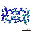
| ファイル | ダウンロード / ファイル: emd_3028.map.gz / 形式: CCP4 / 大きさ: 464.8 KB / タイプ: IMAGE STORED AS FLOATING POINT NUMBER (4 BYTES) | ||||||||||||||||||||||||||||||||||||||||||||||||||||||||||||||||||||
|---|---|---|---|---|---|---|---|---|---|---|---|---|---|---|---|---|---|---|---|---|---|---|---|---|---|---|---|---|---|---|---|---|---|---|---|---|---|---|---|---|---|---|---|---|---|---|---|---|---|---|---|---|---|---|---|---|---|---|---|---|---|---|---|---|---|---|---|---|---|
| 注釈 | microED map of toxic NACore segment, residues 68-78 of alpha-synuclein | ||||||||||||||||||||||||||||||||||||||||||||||||||||||||||||||||||||
| 投影像・断面図 | 画像のコントロール
画像は Spider により作成 これらの図は立方格子座標系で作成されたものです | ||||||||||||||||||||||||||||||||||||||||||||||||||||||||||||||||||||
| ボクセルのサイズ | X: 0.45981 Å / Y: 0.40167 Å / Z: 0.44184 Å | ||||||||||||||||||||||||||||||||||||||||||||||||||||||||||||||||||||
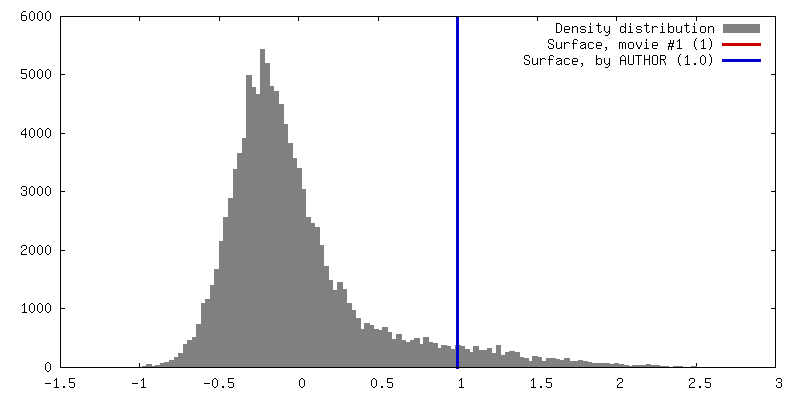
| 密度 |
| ||||||||||||||||||||||||||||||||||||||||||||||||||||||||||||||||||||
| 対称性 | 空間群: 5 | ||||||||||||||||||||||||||||||||||||||||||||||||||||||||||||||||||||
| 詳細 | EMDB XML:
CCP4マップ ヘッダ情報:
| ||||||||||||||||||||||||||||||||||||||||||||||||||||||||||||||||||||
 Y (Sec.)
 X (Row.)
 Z (Col.)


-添付データ
- 試料の構成要素
-全体 : GAVVTGVTAVA, a toxic segment from the NAC domain of Parkinson's d...
| 全体 | 名称: GAVVTGVTAVA, a toxic segment from the NAC domain of Parkinson's disease protein, alpha-synuclein, residues 69-78 |
|---|---|
| 要素 |
|
-超分子 #1000: GAVVTGVTAVA, a toxic segment from the NAC domain of Parkinson's d...
| 超分子 | 名称: GAVVTGVTAVA, a toxic segment from the NAC domain of Parkinson's disease protein, alpha-synuclein, residues 69-78 タイプ: sample / ID: 1000 / 詳細: crystalline fibrils / 集合状態: crystalline fibrils / Number unique components: 1 |
|---|---|
| 分子量 | 理論値: 1 KDa |
-分子 #1: alpha synuclein residues 69-78
| 分子 | 名称: alpha synuclein residues 69-78 / タイプ: protein_or_peptide / ID: 1 / Name.synonym: a-syn 詳細: alpha synuclein residues 68-78. Synthesized chemically. コピー数: 1 / 集合状態: fibril / 組換発現: No / データベース: NCBI |
|---|---|
| 由来(天然) | 生物種: Homo sapiens (ヒト) / 別称: Human |
| 分子量 | 実験値: 1 KDa |
-実験情報
-構造解析
| 手法 | クライオ電子顕微鏡法 |
|---|---|
 解析 解析 | 電子線結晶学 |
| 試料の集合状態 | 3D array |
-試料調製
| 濃度 | 1 mg/mL |
|---|---|
| 緩衝液 | pH: 7 / 詳細: water |
| グリッド | 詳細: quantifoil holey-carbon EM grid, 300 mesh copper |
| 凍結 | 凍結剤: ETHANE / チャンバー内温度: 100 K / 装置: FEI VITROBOT MARK IV 手法: Nanocrystals were deposited onto a quantifoil holey-carbon EM grid in a 2-3 microliter drop after appropriate dilution, which was optimized for crystal density on the grid. All grids were ...手法: Nanocrystals were deposited onto a quantifoil holey-carbon EM grid in a 2-3 microliter drop after appropriate dilution, which was optimized for crystal density on the grid. All grids were then blotted and vitrified by plunging into liquid ethane using a Vitrobot Mark IV (FEI), then transferring to liquid nitrogen for storage. |
| 詳細 | Crystals grew in batch. In a microcentrifuge tube at 37 degrees C with shaking. |
| 結晶化 | 詳細: Crystals grew in batch. In a microcentrifuge tube at 37 degrees C with shaking. |
- 電子顕微鏡法
| 顕微鏡 | FEI TECNAI F20 |
|---|---|
| 温度 | 最低: 99 K / 最高: 101 K / 平均: 100 K |
| 詳細 | very low dose data collection. Spot size 11. |
| 日付 | 2014年8月28日 |
| 撮影 | カテゴリ: CCD フィルム・検出器のモデル: TVIPS TEMCAM-F416 (4k x 4k) 平均電子線量: 0.10000000000000001 e/Å2 / カメラ長: 2230 / 詳細: Diffraction images are available upon request. / ビット/ピクセル: 16 |
| 電子線 | 加速電圧: 200 kV / 電子線源:  FIELD EMISSION GUN FIELD EMISSION GUN |
| 電子光学系 | 照射モード: OTHER / 撮影モード: DIFFRACTION |
| 試料ステージ | 試料ホルダー: liquid nitrogen cooled / 試料ホルダーモデル: GATAN LIQUID NITROGEN / Tilt angle min: -66 / Tilt angle max: 66 / Tilt series - Axis1 - Min angle: -66 ° / Tilt series - Axis1 - Max angle: 66 ° |
| 実験機器 |  モデル: Tecnai F20 / 画像提供: FEI Company |
-画像解析
| 詳細 | Diffraction images were processed with XDS and XSCALE. |
|---|---|
| 最終 再構成 | 解像度のタイプ: BY AUTHOR / 解像度: 1.4 Å / 解像度の算出法: DIFFRACTION PATTERN/LAYERLINES 詳細: The diffraction data set contains intensities measured from four crystals. |
| 結晶パラメータ | 単位格子 - A: 70.81 Å / 単位格子 - B: 4.82 Å / 単位格子 - C: 16.79 Å / 単位格子 - γ: 90.0 ° / 単位格子 - α: 90 ° / 単位格子 - β: 105.68 ° / 空間群: C 1 2 1 |
