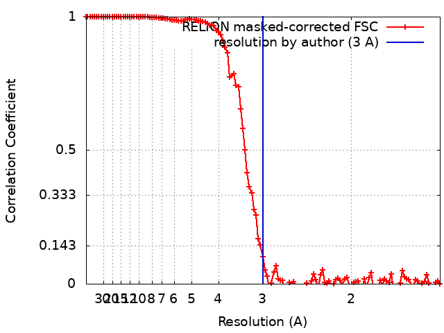
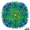
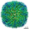
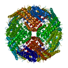
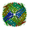
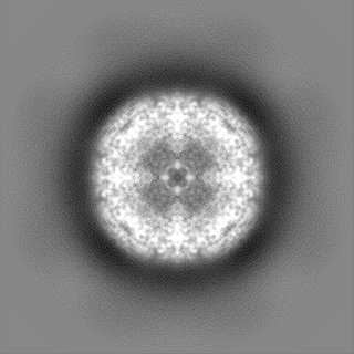
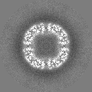
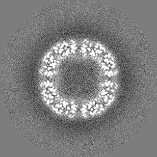
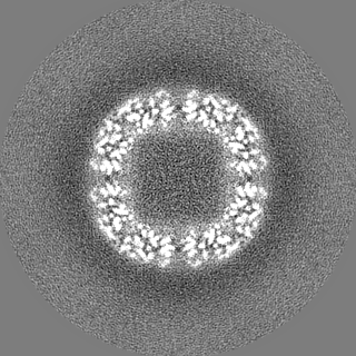
ジャーナル: Sci Rep / 年: 2021 タイトル: Below 3 Å structure of apoferritin using a multipurpose TEM with a side entry cryoholder. 著者: Yoko Kayama / Raymond N Burton-Smith / Chihong Song / Naoya Terahara / Takayuki Kato / Kazuyoshi Murata / 要旨: Recently, the structural analysis of protein complexes by cryo-electron microscopy (cryo-EM) single particle analysis (SPA) has had great impact as a biophysical method. Many results of cryo-EM SPA ...Recently, the structural analysis of protein complexes by cryo-electron microscopy (cryo-EM) single particle analysis (SPA) has had great impact as a biophysical method. Many results of cryo-EM SPA are based on data acquired on state-of-the-art cryo-electron microscopes customized for SPA. These are currently only available in limited locations around the world, where securing machine time is highly competitive. One potential solution for this time-competitive situation is to reuse existing multi-purpose equipment, although this comes with performance limitations. Here, a multi-purpose TEM with a side entry cryo-holder was used to evaluate the potential of high-resolution SPA, resulting in a 3 Å resolution map of apoferritin with local resolution extending to 2.6 Å. This map clearly showed two positions of an aromatic side chain. Further, examination of optimal imaging conditions depending on two different multi-purpose electron microscope and camera combinations was carried out, demonstrating that higher magnifications are not always necessary or desirable. Since automation is effectively a requirement for large-scale data collection, and augmenting the multi-purpose equipment is possible, we expanded testing by acquiring data with SerialEM using a β-galactosidase test sample. This study demonstrates the possibilities of more widely available and established electron microscopes, and their applications for cryo-EM SPA.
 ムービー
ムービー コントローラー


 データを開く
 基本情報
基本情報 マップデータ
 試料
試料 機能・相同性情報
 データ登録者
 引用
引用
 構造の表示
構造の表示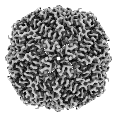
 ダウンロードとリンク
 emd_30096.png
 http://ftp.pdbj.org/pub/emdb/structures/EMD-30096


 Z
Z Y
Y X
X


 試料の構成要素
 解析
解析 電子顕微鏡法
 FIELD EMISSION GUN