 Movie
Movie Controller

[English] 日本語
 Yorodumi
- PDB-7k3v: Apoferritin structure at 1.34 angstrom resolution determined from... -
+ Open data

- Basic information
| Entry | Database: PDB / ID: 7k3v | ||||||||||||
|---|---|---|---|---|---|---|---|---|---|---|---|---|---|
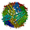
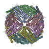
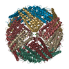
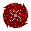
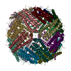
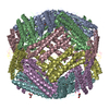
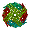
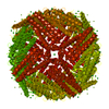
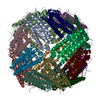
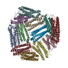
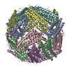
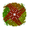
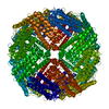
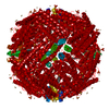
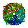
| Title | Apoferritin structure at 1.34 angstrom resolution determined from a 300 kV Titan Krios G3i electron microscope with K3 detector | ||||||||||||
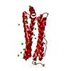
 Components Components | Ferritin heavy chain | ||||||||||||
 Keywords Keywords | OXIDOREDUCTASE / Apoferritin / cryo-EM / atomic resolution / K3 detector | ||||||||||||
| Function / homology |  Function and homology information Function and homology informationiron ion sequestering activity / ferritin complex / Scavenging by Class A Receptors / Golgi Associated Vesicle Biogenesis / ferroxidase / negative regulation of ferroptosis / autolysosome / ferroxidase activity / negative regulation of fibroblast proliferation / ferric iron binding ...iron ion sequestering activity / ferritin complex / Scavenging by Class A Receptors / Golgi Associated Vesicle Biogenesis / ferroxidase / negative regulation of ferroptosis / autolysosome / ferroxidase activity / negative regulation of fibroblast proliferation / ferric iron binding / autophagosome / iron ion transport / ferrous iron binding / Iron uptake and transport / tertiary granule lumen / ficolin-1-rich granule lumen / intracellular iron ion homeostasis / immune response / iron ion binding / negative regulation of cell population proliferation / Neutrophil degranulation / extracellular exosome / extracellular region / identical protein binding / nucleus / cytoplasm / cytosol Similarity search - Function | ||||||||||||
| Biological species |  Homo sapiens (human) Homo sapiens (human) | ||||||||||||
| Method | ELECTRON MICROSCOPY / single particle reconstruction / cryo EM / Resolution: 1.34 Å | ||||||||||||
 Authors Authors | Zhang, K. / Pintilie, G. / Li, S. / Schmid, M. / Chiu, W. | ||||||||||||
| Funding support |  United States, 3items United States, 3items
| ||||||||||||
 Citation Citation | Journal: Cell Res / Year: 2020 Title: Resolving individual atoms of protein complex by cryo-electron microscopy. Authors: Kaiming Zhang / Grigore D Pintilie / Shanshan Li / Michael F Schmid / Wah Chiu / | ||||||||||||
| History |
|
- Structure visualization
| Movie |
Movie viewer |
|---|---|
| Structure viewer | Molecule: MolmilJmol/JSmol |
- Downloads & links
-Download
| PDBx/mmCIF format | 7k3v.cif.gz | 836.3 KB | Display | PDBx/mmCIF format |
|---|---|---|---|---|
| PDB format | pdb7k3v.ent.gz | 687.8 KB | Display | PDB format |
| PDBx/mmJSON format | 7k3v.json.gz | Tree view | PDBx/mmJSON format | |
| Others |  Other downloads Other downloads |
-Validation report
| Arichive directory | https://data.pdbj.org/pub/pdb/validation_reports/k3/7k3vftp://data.pdbj.org/pub/pdb/validation_reports/k3/7k3v | HTTPS FTP |
|---|
-Related structure data
| Related structure data |  22657MC  7k3wC  7rrpC M: map data used to model this data C: citing same article ( |
|---|---|
| Similar structure data |


-Links
 PDBj
PDBj


- Assembly
| Deposited unit | 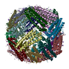
|
|---|---|
| 1 |
|
-Components
| #1: Protein | Mass: 20116.547 Da / Num. of mol.: 24 Source method: isolated from a genetically manipulated source Source: (gene. exp.) Homo sapiens (human) / Gene: FTH1, FTH, FTHL6, OK/SW-cl.84, PIG15Production host:  References: UniProt: P02794, ferroxidase #2: Chemical | ChemComp-ZN /   Mass: 65.409 Da / Num. of mol.: 254 / Source method: obtained synthetically / Formula: Zn Mass: 65.409 Da / Num. of mol.: 254 / Source method: obtained synthetically / Formula: Zn#3: Chemical | ChemComp-NA /   Mass: 22.990 Da / Num. of mol.: 80 / Source method: obtained synthetically / Formula: Na Mass: 22.990 Da / Num. of mol.: 80 / Source method: obtained synthetically / Formula: Na#4: Water | ChemComp-HOH / |  Mass: 18.015 Da / Num. of mol.: 2672 / Source method: isolated from a natural source / Formula: H2O Mass: 18.015 Da / Num. of mol.: 2672 / Source method: isolated from a natural source / Formula: H2OHas ligand of interest | N | |
|---|
-Experimental details
-Experiment
| Experiment | Method: ELECTRON MICROSCOPY |
|---|---|
| EM experiment | Aggregation state: PARTICLE / 3D reconstruction method: single particle reconstruction |
- Sample preparation
| Component | Name: Apoferritin / Type: COMPLEX / Entity ID: #1 / Source: RECOMBINANT |
|---|---|
| Molecular weight | Value: 0.48 MDa / Experimental value: YES |
| Source (natural) | Organism: Homo sapiens (human) |
| Source (recombinant) | Organism: |
| Buffer solution | pH: 8 / Details: 50 mM Tris-HCl (pH 8.0), 150 mM NaCl |
| Specimen | Conc.: 1.5 mg/ml / Embedding applied: NO / Shadowing applied: NO / Staining applied: NO / Vitrification applied: YES |
| Specimen support | Grid material: COPPER / Grid mesh size: 200 divisions/in. / Grid type: Quantifoil R2/1 |
| Vitrification | Cryogen name: ETHANE |
- Electron microscopy imaging
| Experimental equipment |  Model: Titan Krios / Image courtesy: FEI Company |
|---|---|
| Microscopy | Model: FEI TITAN KRIOS |
| Electron gun | Electron source:  FIELD EMISSION GUN / Accelerating voltage: 300 kV / Illumination mode: FLOOD BEAM FIELD EMISSION GUN / Accelerating voltage: 300 kV / Illumination mode: FLOOD BEAM |
| Electron lens | Mode: BRIGHT FIELD / Nominal defocus max: 1000 nm / Nominal defocus min: 300 nm / Calibrated defocus min: 100 nm / Calibrated defocus max: 1500 nm / Cs: 2.7 mm / C2 aperture diameter: 70 µm / Alignment procedure: COMA FREE |
| Specimen holder | Cryogen: NITROGEN / Specimen holder model: FEI TITAN KRIOS AUTOGRID HOLDER |
| Image recording | Average exposure time: 0.5 sec. / Electron dose: 45 e/Å2 / Film or detector model: GATAN K3 BIOQUANTUM (6k x 4k) / Num. of grids imaged: 1 / Num. of real images: 8034 |
| EM imaging optics | Energyfilter name: GIF Bioquantum / Energyfilter slit width: 15 eV |
- Processing
| Software | Name: Segger / Version: 2.5.2 / Classification: refinement | |||||||||||||||||||||||||||||||||||
|---|---|---|---|---|---|---|---|---|---|---|---|---|---|---|---|---|---|---|---|---|---|---|---|---|---|---|---|---|---|---|---|---|---|---|---|---|
| EM software |
| |||||||||||||||||||||||||||||||||||
| CTF correction | Type: PHASE FLIPPING AND AMPLITUDE CORRECTION | |||||||||||||||||||||||||||||||||||
| Particle selection | Num. of particles selected: 1176336 | |||||||||||||||||||||||||||||||||||
| Symmetry | Point symmetry: O (octahedral) | |||||||||||||||||||||||||||||||||||
| 3D reconstruction | Resolution: 1.34 Å / Resolution method: FSC 0.143 CUT-OFF / Num. of particles: 902455 / Symmetry type: POINT | |||||||||||||||||||||||||||||||||||
| Atomic model building | Protocol: OTHER / Space: REAL / Target criteria: CC | |||||||||||||||||||||||||||||||||||
| Atomic model building | PDB-ID: 3AJO Pdb chain-ID: A / Accession code: 3AJO / Source name: PDB / Type: experimental model |

