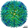
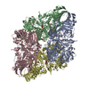
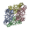
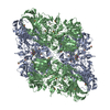
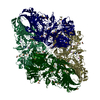
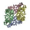
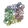
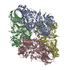
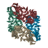
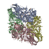
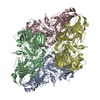
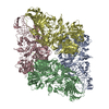
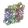
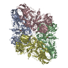
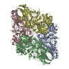
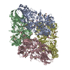
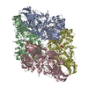
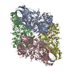
Journal: Sci Rep / Year: 2021 Title: Below 3 Å structure of apoferritin using a multipurpose TEM with a side entry cryoholder. Authors: Yoko Kayama / Raymond N Burton-Smith / Chihong Song / Naoya Terahara / Takayuki Kato / Kazuyoshi Murata / Abstract: Recently, the structural analysis of protein complexes by cryo-electron microscopy (cryo-EM) single particle analysis (SPA) has had great impact as a biophysical method. Many results of cryo-EM SPA ...Recently, the structural analysis of protein complexes by cryo-electron microscopy (cryo-EM) single particle analysis (SPA) has had great impact as a biophysical method. Many results of cryo-EM SPA are based on data acquired on state-of-the-art cryo-electron microscopes customized for SPA. These are currently only available in limited locations around the world, where securing machine time is highly competitive. One potential solution for this time-competitive situation is to reuse existing multi-purpose equipment, although this comes with performance limitations. Here, a multi-purpose TEM with a side entry cryo-holder was used to evaluate the potential of high-resolution SPA, resulting in a 3 Å resolution map of apoferritin with local resolution extending to 2.6 Å. This map clearly showed two positions of an aromatic side chain. Further, examination of optimal imaging conditions depending on two different multi-purpose electron microscope and camera combinations was carried out, demonstrating that higher magnifications are not always necessary or desirable. Since automation is effectively a requirement for large-scale data collection, and augmenting the multi-purpose equipment is possible, we expanded testing by acquiring data with SerialEM using a β-galactosidase test sample. This study demonstrates the possibilities of more widely available and established electron microscopes, and their applications for cryo-EM SPA.
History
Deposition
Mar 12, 2020
-
Header (metadata) release
Mar 17, 2021
-
Map release
Mar 17, 2021
-
Update
Mar 17, 2021
-
Current status
Mar 17, 2021
Processing site: PDBj / Status: Released
-
Structure visualization
Movie
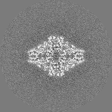
Surface view with section colored by density value
In the structure databanks used in Yorodumi, some data are registered as the other names, "COVID-19 virus" and "2019-nCoV". Here are the details of the virus and the list of structure data.
Jan 31, 2019. EMDB accession codes are about to change! (news from PDBe EMDB page)
EMDB accession codes are about to change! (news from PDBe EMDB page)
The allocation of 4 digits for EMDB accession codes will soon come to an end. Whilst these codes will remain in use, new EMDB accession codes will include an additional digit and will expand incrementally as the available range of codes is exhausted. The current 4-digit format prefixed with “EMD-” (i.e. EMD-XXXX) will advance to a 5-digit format (i.e. EMD-XXXXX), and so on. It is currently estimated that the 4-digit codes will be depleted around Spring 2019, at which point the 5-digit format will come into force.
The EM Navigator/Yorodumi systems omit the EMD- prefix.
Related info.:Q: What is EMD? / ID/Accession-code notation in Yorodumi/EM Navigator
Yorodumi is a browser for structure data from EMDB, PDB, SASBDB, etc.
This page is also the successor to EM Navigator detail page, and also detail information page/front-end page for Omokage search.
The word "yorodu" (or yorozu) is an old Japanese word meaning "ten thousand". "mi" (miru) is to see.
Related info.:EMDB / PDB / SASBDB / Comparison of 3 databanks / Yorodumi Search / Aug 31, 2016. New EM Navigator & Yorodumi / Yorodumi Papers / Jmol/JSmol / Function and homology information / Changes in new EM Navigator and Yorodumi
 Movie
Movie Controller

 Yorodumi
 Open data
 Basic information
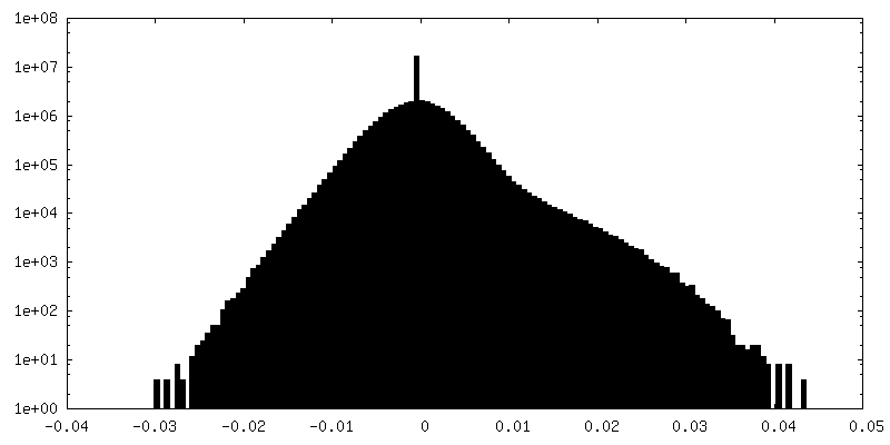
 Map data
 Sample
 Function and homology information
 Authors
 Citation
 Structure visualization
 Downloads & links
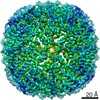
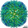
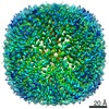
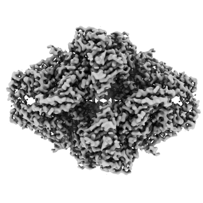 emd_30095.png
 http://ftp.pdbj.org/pub/emdb/structures/EMD-30095


 Z (Sec.)
 Y (Row.)
 X (Col.)


 Sample components
 Processing
 Electron microscopy
 FIELD EMISSION GUN
