 Movie
Movie Controller


+ Open data

- Basic information
| Entry | 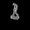 | |||||||||
|---|---|---|---|---|---|---|---|---|---|---|
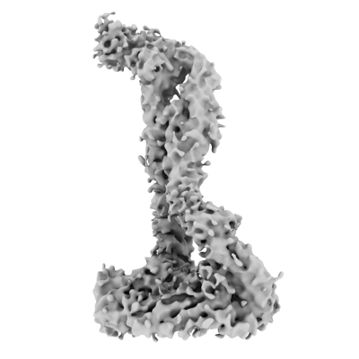
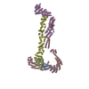
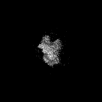
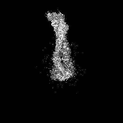
| Title | Consensus Map of the Dsl1:Qb:Qc Complex | |||||||||
 Map data Map data | Sharpened map, Consensus Map, of the Dsl1:Qb:Qc complex | |||||||||
 Sample Sample |
| |||||||||
 Keywords Keywords | Tether / SNARE / Complex / TRANSPORT PROTEIN | |||||||||
| Biological species |  | |||||||||
| Method | single particle reconstruction / cryo EM / Resolution: 6.2 Å | |||||||||
 Authors Authors | DAmico KA / Jeffrey PD / Hughson FM | |||||||||
| Funding support |  United States, 2 items United States, 2 items
| |||||||||
 Citation Citation | Journal: TO BE PUBLISHED Title: CryoEM structure of the Dsl1 complex bound to SNAREs Sec20 and Use1 Authors: DAmico KA / Stanton AE / Shirkey JD / Jeffrey PD / Hughson FM | |||||||||
| History |
|
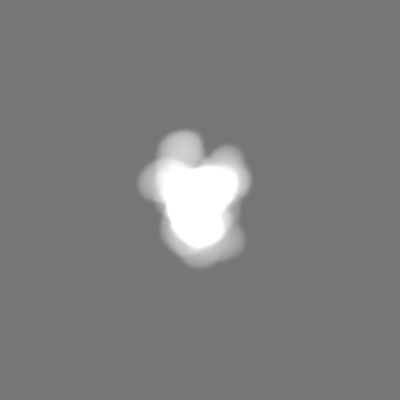
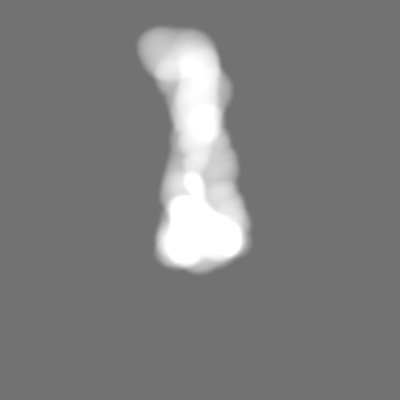
- Structure visualization
| Supplemental images |
|---|
- Downloads & links
-EMDB archive
| Map data | emd_28774.map.gz | 229.9 MB |  EMDB map data format EMDB map data format | |
|---|---|---|---|---|
| Header (meta data) | emd-28774-v30.xmlemd-28774.xml | 18.6 KB 18.6 KB | Display Display | EMDB header |
| FSC (resolution estimation) | emd_28774_fsc.xml | 13.3 KB | Display | FSC data file |
| Images |  emd_28774.png emd_28774.png | 60.7 KB | ||
| Masks | emd_28774_msk_1.map | 244.1 MB | Mask map | |
| Filedesc metadata | emd-28774.cif.gz | 4.8 KB | ||
| Others | emd_28774_additional_1.map.gzemd_28774_half_map_1.map.gzemd_28774_half_map_2.map.gz | 120.3 MB 226.9 MB 226.9 MB | ||
| Archive directory |  http://ftp.pdbj.org/pub/emdb/structures/EMD-28774ftp://ftp.pdbj.org/pub/emdb/structures/EMD-28774 http://ftp.pdbj.org/pub/emdb/structures/EMD-28774ftp://ftp.pdbj.org/pub/emdb/structures/EMD-28774 | HTTPS FTP |
-Validation report
| Summary document | emd_28774_validation.pdf.gz | 945.1 KB | Display | EMDB validaton report |
|---|---|---|---|---|
| Full document | emd_28774_full_validation.pdf.gz | 944.6 KB | Display | |
| Data in XML | emd_28774_validation.xml.gz | 22.3 KB | Display | |
| Data in CIF | emd_28774_validation.cif.gz | 28.7 KB | Display | |
| Arichive directory | https://ftp.pdbj.org/pub/emdb/validation_reports/EMD-28774ftp://ftp.pdbj.org/pub/emdb/validation_reports/EMD-28774 | HTTPS FTP |
-Related structure data
-Links
| EMDB pages | EMDB (EBI/PDBe) / EMDataResource |
|---|
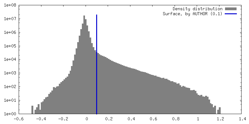
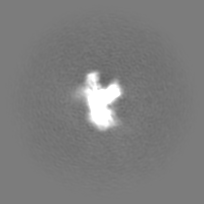
-Map
| File | Download / File: emd_28774.map.gz / Format: CCP4 / Size: 244.1 MB / Type: IMAGE STORED AS FLOATING POINT NUMBER (4 BYTES) | ||||||||||||||||||||||||||||||||||||
|---|---|---|---|---|---|---|---|---|---|---|---|---|---|---|---|---|---|---|---|---|---|---|---|---|---|---|---|---|---|---|---|---|---|---|---|---|---|
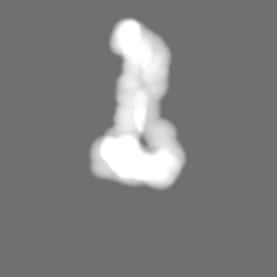
| Annotation | Sharpened map, Consensus Map, of the Dsl1:Qb:Qc complex | ||||||||||||||||||||||||||||||||||||
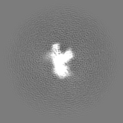
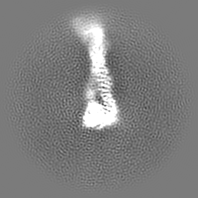
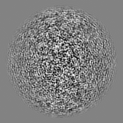
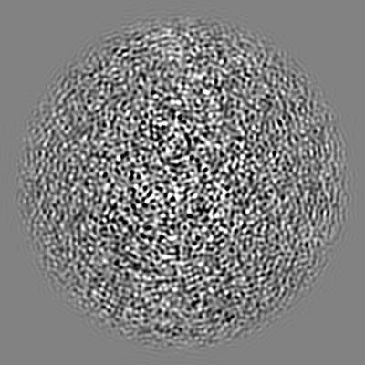
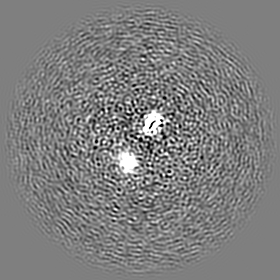
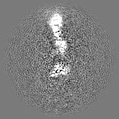
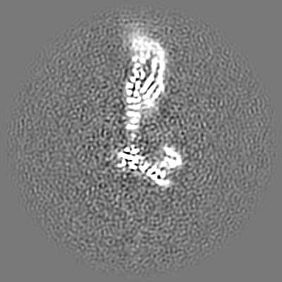
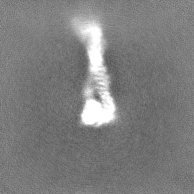
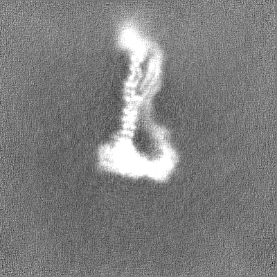
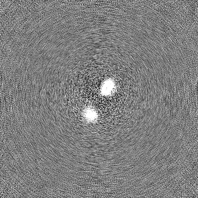
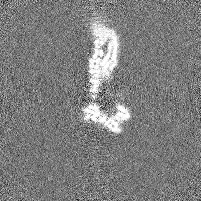
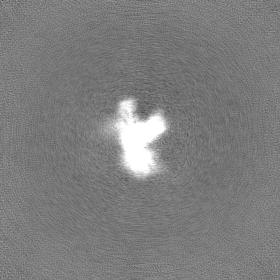
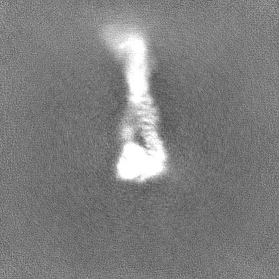
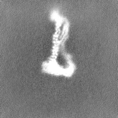
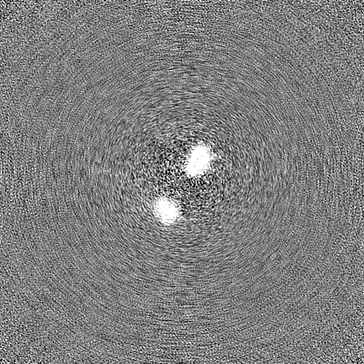
| Projections & slices | Image control
Images are generated by Spider. | ||||||||||||||||||||||||||||||||||||
| Voxel size | X=Y=Z: 1.114 Å | ||||||||||||||||||||||||||||||||||||
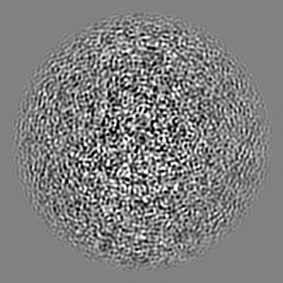
| Density |
| ||||||||||||||||||||||||||||||||||||
| Symmetry | Space group: 1 | ||||||||||||||||||||||||||||||||||||
| Details | EMDB XML:
|
 Z (Sec.)
 Y (Row.)
 X (Col.)


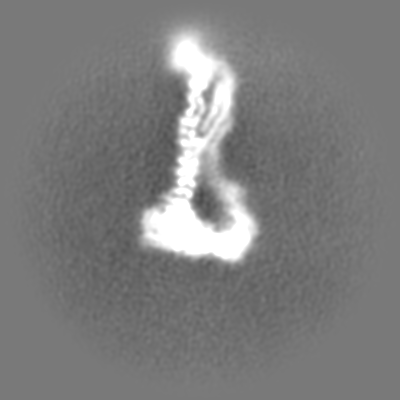
-Supplemental data
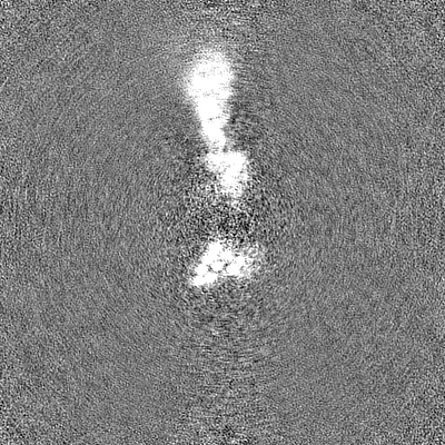
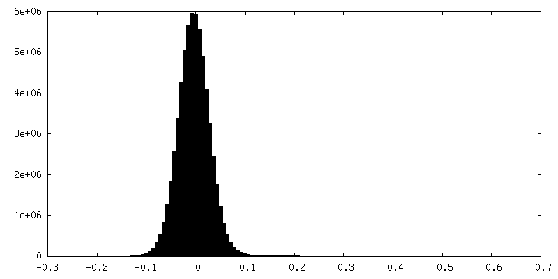
-Mask #1
| File | emd_28774_msk_1.map | ||||||||||||
|---|---|---|---|---|---|---|---|---|---|---|---|---|---|
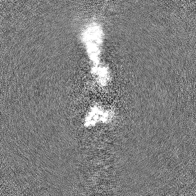
| Projections & Slices |
| ||||||||||||
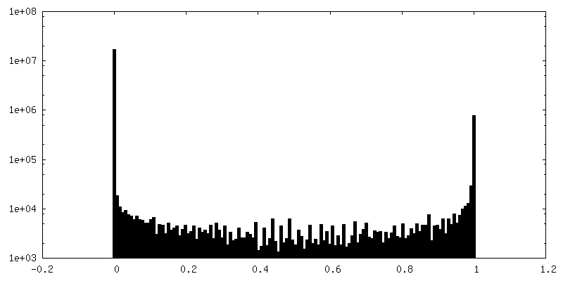
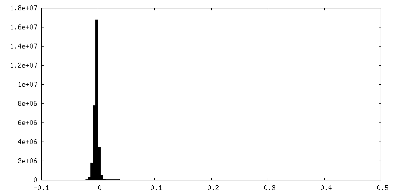
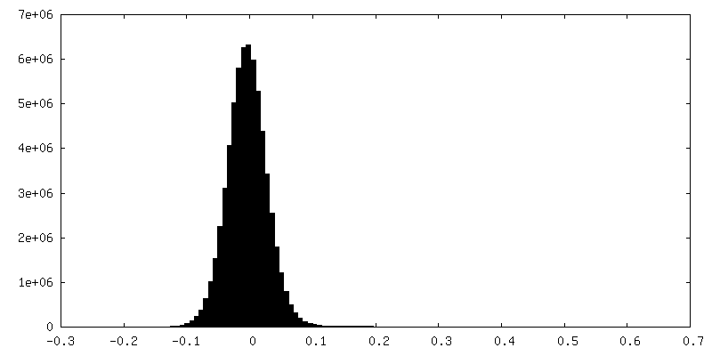
| Density Histograms |


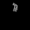
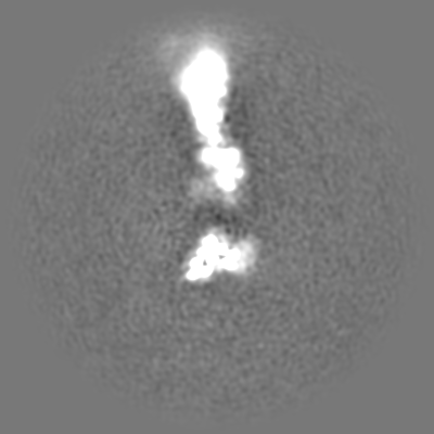
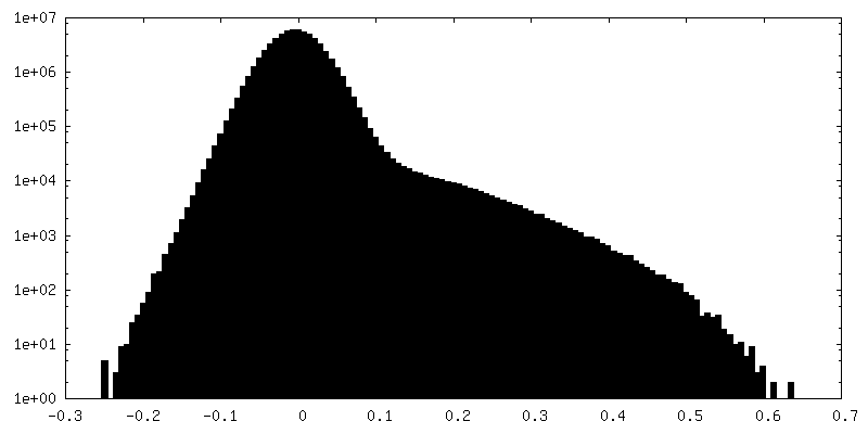
-Additional map: Unsharpened map, Consensus Map, of the Dsl1:Qb:Qc complex
| File | emd_28774_additional_1.map | ||||||||||||
|---|---|---|---|---|---|---|---|---|---|---|---|---|---|
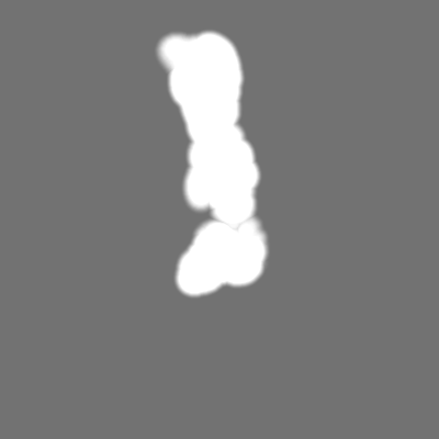
| Annotation | Unsharpened map, Consensus Map, of the Dsl1:Qb:Qc complex | ||||||||||||
| Projections & Slices |
| ||||||||||||
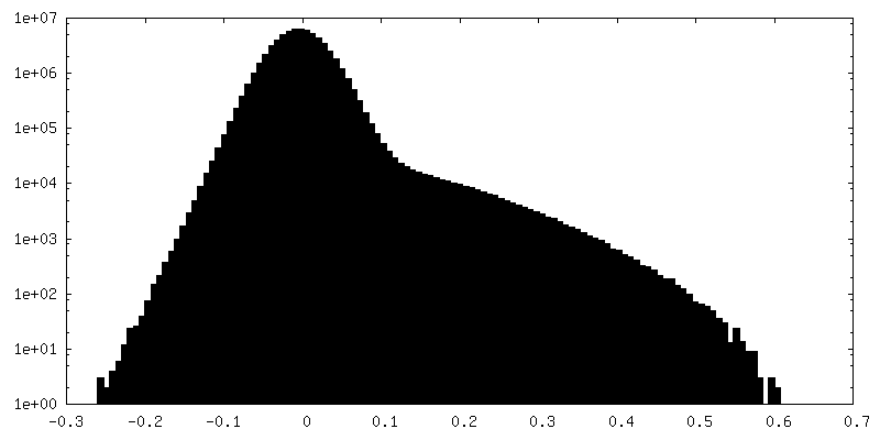
| Density Histograms |


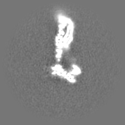

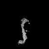
-Half map: Half map B, Consensus Map, of the Dsl1:Qb:Qc complex
| File | emd_28774_half_map_1.map | ||||||||||||
|---|---|---|---|---|---|---|---|---|---|---|---|---|---|
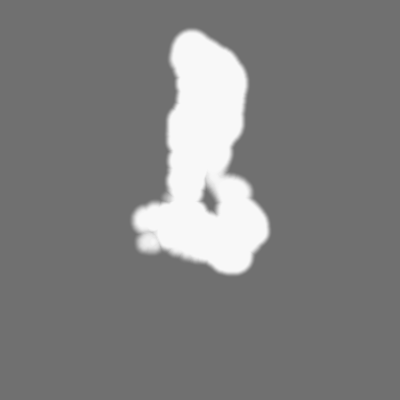
| Annotation | Half map B, Consensus Map, of the Dsl1:Qb:Qc complex | ||||||||||||
| Projections & Slices |
| ||||||||||||
| Density Histograms |

-Half map: Half map A, Consensus Map, of the Dsl1:Qb:Qc complex
| File | emd_28774_half_map_2.map | ||||||||||||
|---|---|---|---|---|---|---|---|---|---|---|---|---|---|
| Annotation | Half map A, Consensus Map, of the Dsl1:Qb:Qc complex | ||||||||||||
| Projections & Slices |
| ||||||||||||
| Density Histograms |

- Sample components
-Entire : Dsl1 complex bound to SNARE proteins Sec20 and Use1
| Entire | Name: Dsl1 complex bound to SNARE proteins Sec20 and Use1 |
|---|---|
| Components |
|
-Supramolecule #1: Dsl1 complex bound to SNARE proteins Sec20 and Use1
| Supramolecule | Name: Dsl1 complex bound to SNARE proteins Sec20 and Use1 / type: complex / ID: 1 / Parent: 0 |
|---|---|
| Source (natural) | Organism: |
| Molecular weight | Theoretical: 255.41261 KDa |
-Supramolecule #2: Dsl1 Complex
| Supramolecule | Name: Dsl1 Complex / type: complex / ID: 2 / Parent: 1 |
|---|---|
| Source (natural) | Organism: |
-Experimental details
-Structure determination
| Method | cryo EM |
|---|---|
 Processing Processing | single particle reconstruction |
| Aggregation state | particle |
-Sample preparation
| Concentration | 3 mg/mL | |||||||||||||||
|---|---|---|---|---|---|---|---|---|---|---|---|---|---|---|---|---|
| Buffer | pH: 7.5 Component:
Details: Buffer was made fresh from concentrated components and sterile filtered. NP40 was not present during protein purification but was an additive during the grid preparation. | |||||||||||||||
| Grid | Model: Quantifoil R1.2/1.3 / Material: COPPER / Mesh: 300 / Support film - Material: CARBON / Support film - topology: HOLEY / Support film - Film thickness: 10 | |||||||||||||||
| Vitrification | Cryogen name: ETHANE / Chamber humidity: 100 % / Chamber temperature: 277.15 K / Instrument: FEI VITROBOT MARK IV / Details: Force=0 Wait Time=0 Blot Time=6s Drain Time=0. | |||||||||||||||
| Details | Sample was consistently in the thickest regions of ice only, often close to the edges of the carbon hole |
- Electron microscopy
| Microscope | FEI TITAN KRIOS |
|---|---|
| Image recording | Film or detector model: GATAN K2 SUMMIT (4k x 4k) / Number grids imaged: 1 / Number real images: 5857 / Average electron dose: 45.0 e/Å2 |
| Electron beam | Acceleration voltage: 300 kV / Electron source:  FIELD EMISSION GUN FIELD EMISSION GUN |
| Electron optics | Illumination mode: FLOOD BEAM / Imaging mode: BRIGHT FIELD / Nominal defocus max: 2.5 µm / Nominal defocus min: 1.25 µm |
| Experimental equipment |  Model: Titan Krios / Image courtesy: FEI Company |

