Movie
Movie Controller

[English] 日本語
 Yorodumi
- EMDB-2787: The structure of the large subunit of the mammalian mitoribosome -
+ Open data

- Basic information
| Entry | Database: EMDB / ID: EMD-2787 | |||||||||
|---|---|---|---|---|---|---|---|---|---|---|
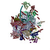
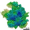
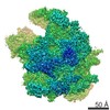
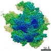
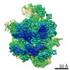
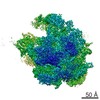
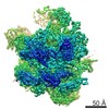
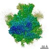
| Title | The structure of the large subunit of the mammalian mitoribosome | |||||||||
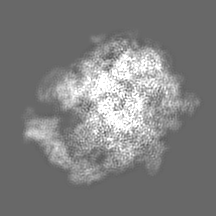
 Map data Map data | Reconstruction of the mammalian mitoribosomal 39S subunit at 3.4 Angstrom resolution | |||||||||
 Sample Sample |
| |||||||||
 Keywords Keywords | mammalian mitochondrial ribosome / 39S large ribosomal subunit / translation / ribosomal proteins / rRNA / tRNA / polypeptide exit site / peptidyl transferase center | |||||||||
| Function / homology |  Function and homology information Function and homology informationMitochondrial translation elongation / Mitochondrial translation termination / rRNA import into mitochondrion / mitochondrial translational termination / mitochondrial translational elongation / ribonuclease III activity / translation release factor activity, codon nonspecific / Mitochondrial protein degradation / mitochondrial large ribosomal subunit / peptidyl-tRNA hydrolase ...Mitochondrial translation elongation / Mitochondrial translation termination / rRNA import into mitochondrion / mitochondrial translational termination / mitochondrial translational elongation / ribonuclease III activity / translation release factor activity, codon nonspecific / Mitochondrial protein degradation / mitochondrial large ribosomal subunit / peptidyl-tRNA hydrolase / mitochondrial ribosome / peptidyl-tRNA hydrolase activity / mitochondrial translation / organelle membrane / RNA processing / cell junction / large ribosomal subunit / double-stranded RNA binding / 5S rRNA binding / rRNA binding / nuclear body / structural constituent of ribosome / ribosome / translation / ribonucleoprotein complex / protein domain specific binding / nucleotide binding / intracellular membrane-bounded organelle / mitochondrion / RNA binding / nucleoplasm / nucleus / plasma membrane / cytoplasm / cytosol Similarity search - Function | |||||||||
| Biological species |  | |||||||||
| Method | single particle reconstruction / cryo EM / Resolution: 3.4 Å | |||||||||
 Authors Authors | Greber BJ / Boehringer D / Leibundgut M / Bieri P / Leitner A / Schmitz N / Aebersold R / Ban N | |||||||||
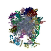
 Citation Citation | Journal: Nature / Year: 2014 Title: The complete structure of the large subunit of the mammalian mitochondrial ribosome. Authors: Basil J Greber / Daniel Boehringer / Marc Leibundgut / Philipp Bieri / Alexander Leitner / Nikolaus Schmitz / Ruedi Aebersold / Nenad Ban /  Abstract: Mitochondrial ribosomes (mitoribosomes) are extensively modified ribosomes of bacterial descent specialized for the synthesis and insertion of membrane proteins that are critical for energy ...Mitochondrial ribosomes (mitoribosomes) are extensively modified ribosomes of bacterial descent specialized for the synthesis and insertion of membrane proteins that are critical for energy conversion and ATP production inside mitochondria. Mammalian mitoribosomes, which comprise 39S and 28S subunits, have diverged markedly from the bacterial ribosomes from which they are derived, rendering them unique compared to bacterial, eukaryotic cytosolic and fungal mitochondrial ribosomes. We have previously determined at 4.9 Å resolution the architecture of the porcine (Sus scrofa) 39S subunit, which is highly homologous to the human mitoribosomal large subunit. Here we present the complete atomic structure of the porcine 39S large mitoribosomal subunit determined in the context of a stalled translating mitoribosome at 3.4 Å resolution by cryo-electron microscopy and chemical crosslinking/mass spectrometry. The structure reveals the locations and the detailed folds of 50 mitoribosomal proteins, shows the highly conserved mitoribosomal peptidyl transferase active site in complex with its substrate transfer RNAs, and defines the path of the nascent chain in mammalian mitoribosomes along their idiosyncratic exit tunnel. Furthermore, we present evidence that a mitochondrial tRNA has become an integral component of the central protuberance of the 39S subunit where it architecturally substitutes for the absence of the 5S ribosomal RNA, a ubiquitous component of all cytoplasmic ribosomes. | |||||||||
| History |
|
- Structure visualization
| Movie |
Movie viewer |
|---|---|
| Structure viewer | EM map: SurfViewMolmilJmol/JSmol |
| Supplemental images |

- Downloads & links
-EMDB archive
| Map data | emd_2787.map.gz | 6.9 MB | EMDB map data format | |
|---|---|---|---|---|
| Header (meta data) | emd-2787-v30.xmlemd-2787.xml | 11.6 KB 11.6 KB | Display Display | EMDB header |
| Images | 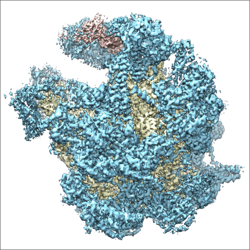 EMD-2787-image.png EMD-2787-image.png | 352.7 KB | ||
| Archive directory |  http://ftp.pdbj.org/pub/emdb/structures/EMD-2787ftp://ftp.pdbj.org/pub/emdb/structures/EMD-2787 http://ftp.pdbj.org/pub/emdb/structures/EMD-2787ftp://ftp.pdbj.org/pub/emdb/structures/EMD-2787 | HTTPS FTP |
-Validation report
| Summary document | emd_2787_validation.pdf.gz | 267 KB | Display | EMDB validaton report |
|---|---|---|---|---|
| Full document | emd_2787_full_validation.pdf.gz | 266.1 KB | Display | |
| Data in XML | emd_2787_validation.xml.gz | 5.8 KB | Display | |
| Arichive directory | https://ftp.pdbj.org/pub/emdb/validation_reports/EMD-2787ftp://ftp.pdbj.org/pub/emdb/validation_reports/EMD-2787 | HTTPS FTP |
-Related structure data
| Related structure data | 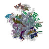 4v19MC 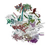 4v1aMC M: atomic model generated by this map C: citing same article ( |
|---|---|
| Similar structure data |


-Links
| EMDB pages | EMDB (EBI/PDBe) / EMDataResource |
|---|---|
| Related items in Molecule of the Month |


-Map
| File | Download / File: emd_2787.map.gz / Format: CCP4 / Size: 37.5 MB / Type: IMAGE STORED AS FLOATING POINT NUMBER (4 BYTES) | ||||||||||||||||||||||||||||||||||||||||||||||||||||||||||||||||||||
|---|---|---|---|---|---|---|---|---|---|---|---|---|---|---|---|---|---|---|---|---|---|---|---|---|---|---|---|---|---|---|---|---|---|---|---|---|---|---|---|---|---|---|---|---|---|---|---|---|---|---|---|---|---|---|---|---|---|---|---|---|---|---|---|---|---|---|---|---|---|
| Annotation | Reconstruction of the mammalian mitoribosomal 39S subunit at 3.4 Angstrom resolution | ||||||||||||||||||||||||||||||||||||||||||||||||||||||||||||||||||||
| Projections & slices | Image control
Images are generated by Spider. | ||||||||||||||||||||||||||||||||||||||||||||||||||||||||||||||||||||
| Voxel size | X=Y=Z: 1.4 Å | ||||||||||||||||||||||||||||||||||||||||||||||||||||||||||||||||||||
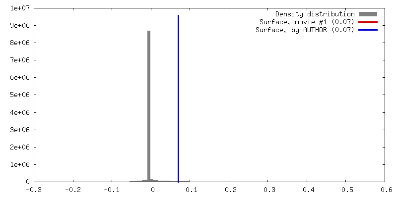
| Density |
| ||||||||||||||||||||||||||||||||||||||||||||||||||||||||||||||||||||
| Symmetry | Space group: 1 | ||||||||||||||||||||||||||||||||||||||||||||||||||||||||||||||||||||
| Details | EMDB XML:
CCP4 map header:
| ||||||||||||||||||||||||||||||||||||||||||||||||||||||||||||||||||||
 Z (Sec.)
 Y (Row.)
 X (Col.)


-Supplemental data
- Sample components
-Entire : mammalian mitochondrial ribosome
| Entire | Name: mammalian mitochondrial ribosome |
|---|---|
| Components |
|
-Supramolecule #1000: mammalian mitochondrial ribosome
| Supramolecule | Name: mammalian mitochondrial ribosome / type: sample / ID: 1000 / Oligomeric state: monomer / Number unique components: 1 |
|---|---|
| Molecular weight | Theoretical: 2.7 MDa |
-Supramolecule #1: 39S large subunit of the mitochondrial ribosome
| Supramolecule | Name: 39S large subunit of the mitochondrial ribosome / type: organelle_or_cellular_component / ID: 1 / Number of copies: 1 / Oligomeric state: Monomer / Recombinant expression: No |
|---|---|
| Ref GO | 0: GO:0005761 |
| Source (natural) | Organism: |
| Molecular weight | Theoretical: 1.6 MDa |
-Experimental details
-Structure determination
| Method | cryo EM |
|---|---|
 Processing Processing | single particle reconstruction |
| Aggregation state | particle |
-Sample preparation
| Buffer | pH: 7.4 / Details: 20 mM HEPES-KOH, 50 mM KCl, 1 mM DTT, 40 mM MgCl2 |
|---|---|
| Grid | Details: 200 mesh Quantifoil R 2/2 holey carbon grids with a thin continuous carbon support film applied |
| Vitrification | Cryogen name: ETHANE-PROPANE MIXTURE / Instrument: HOMEMADE PLUNGER |
- Electron microscopy #1
| Microscopy ID | 1 |
|---|---|
| Microscope | FEI TITAN KRIOS |
| Details | single movie frame readout: 7 frames per exposure |
| Date | May 3, 2014 |
| Image recording | Category: CCD / Film or detector model: FEI FALCON II (4k x 4k) / Digitization - Sampling interval: 14 µm / Average electron dose: 20 e/Å2 Details: individual movie frame readout: 7 frames per exposure |
| Electron beam | Acceleration voltage: 300 kV / Electron source:  FIELD EMISSION GUN FIELD EMISSION GUN |
| Electron optics | Calibrated magnification: 100000 / Illumination mode: FLOOD BEAM / Imaging mode: BRIGHT FIELD / Cs: 2.7 mm / Nominal defocus max: 3.0 µm / Nominal defocus min: 0.8 µm / Nominal magnification: 59000 |
| Sample stage | Specimen holder model: FEI TITAN KRIOS AUTOGRID HOLDER |
| Experimental equipment |  Model: Titan Krios / Image courtesy: FEI Company |
-Electron microscopy #2
| Microscopy ID | 2 |
|---|---|
| Microscope | FEI TITAN KRIOS |
| Details | single movie frame readout: 7 frames per exposure |
| Date | May 30, 2014 |
| Image recording | Category: CCD / Film or detector model: FEI FALCON II (4k x 4k) / Digitization - Sampling interval: 14 µm / Average electron dose: 20 e/Å2 Details: individual movie frame readout: 7 frames per exposure |
| Electron beam | Acceleration voltage: 300 kV / Electron source: FIELD EMISSION GUN |
| Electron optics | Calibrated magnification: 100000 / Illumination mode: FLOOD BEAM / Imaging mode: BRIGHT FIELD / Cs: 2.7 mm / Nominal defocus max: 3.0 µm / Nominal defocus min: 0.8 µm / Nominal magnification: 59000 |
| Sample stage | Specimen holder model: FEI TITAN KRIOS AUTOGRID HOLDER |
| Experimental equipment | Model: Titan Krios / Image courtesy: FEI Company |
-Image processing
| Details | Particles were selected in batchboxer (EMAN 1.9) and extracted using RELION 1.2. |
|---|---|
| CTF correction | Details: per detector frame |
| Final reconstruction | Applied symmetry - Point group: C1 (asymmetric) / Algorithm: OTHER / Resolution.type: BY AUTHOR / Resolution: 3.4 Å / Resolution method: OTHER / Software - Name: CTFFIND3, RELION, 1.2 / Number images used: 141675 |
-Atomic model buiding 1
| Initial model | PDB ID: |
|---|---|
| Software | Name: Chimera, O, COOT |
| Details | The initial coordinate model was fitted into the cryo-EM density using Chimera. The model was then extended and completely rebuilt using COOT (RNA) and O (proteins). |
| Refinement | Space: REAL / Protocol: RIGID BODY FIT |
| Output model | PDB-4v19: PDB-4v1a: |