 Movie
Movie Controller

[English] 日本語
 Yorodumi
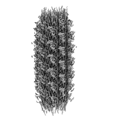
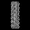
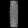
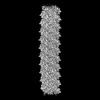
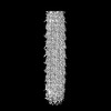
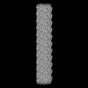
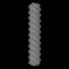
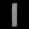
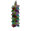
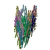
- EMDB-27059: Cryo-EM structure of the helical E. coli K12 flagellar filament core -
+ Open data

- Basic information
| Entry | 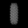 | |||||||||
|---|---|---|---|---|---|---|---|---|---|---|
| Title | Cryo-EM structure of the helical E. coli K12 flagellar filament core | |||||||||
 Map data Map data | Cryo-EM structure of the helical E. coli K12 flagellar filament core | |||||||||
 Sample Sample |
| |||||||||
| Biological species |  | |||||||||
| Method | helical reconstruction / cryo EM / Resolution: 3.0 Å | |||||||||
 Authors Authors | Sonani RR / Kreutzberger MAB / Sebastian AL / Scharf B / Egelman EH | |||||||||
| Funding support |  United States, 1 items United States, 1 items
| |||||||||
 Citation Citation | Journal: Cell / Year: 2022 Title: Convergent evolution in the supercoiling of prokaryotic flagellar filaments. Authors: Mark A B Kreutzberger / Ravi R Sonani / Junfeng Liu / Sharanya Chatterjee / Fengbin Wang / Amanda L Sebastian / Priyanka Biswas / Cheryl Ewing / Weili Zheng / Frédéric Poly / Gad Frankel / ...Authors: Mark A B Kreutzberger / Ravi R Sonani / Junfeng Liu / Sharanya Chatterjee / Fengbin Wang / Amanda L Sebastian / Priyanka Biswas / Cheryl Ewing / Weili Zheng / Frédéric Poly / Gad Frankel / B F Luisi / Chris R Calladine / Mart Krupovic / Birgit E Scharf / Edward H Egelman /   Abstract: The supercoiling of bacterial and archaeal flagellar filaments is required for motility. Archaeal flagellar filaments have no homology to their bacterial counterparts and are instead homologs of ...The supercoiling of bacterial and archaeal flagellar filaments is required for motility. Archaeal flagellar filaments have no homology to their bacterial counterparts and are instead homologs of bacterial type IV pili. How these prokaryotic flagellar filaments, each composed of thousands of copies of identical subunits, can form stable supercoils under torsional stress is a fascinating puzzle for which structural insights have been elusive. Advances in cryoelectron microscopy (cryo-EM) make it now possible to directly visualize the basis for supercoiling, and here, we show the atomic structures of supercoiled bacterial and archaeal flagellar filaments. For the bacterial flagellar filament, we identify 11 distinct protofilament conformations with three broad classes of inter-protomer interface. For the archaeal flagellar filament, 10 protofilaments form a supercoil geometry supported by 10 distinct conformations, with one inter-protomer discontinuity creating a seam inside of the curve. Our results suggest that convergent evolution has yielded stable superhelical geometries that enable microbial locomotion. | |||||||||
| History |
|
- Structure visualization
| Supplemental images |
|---|
- Downloads & links
-EMDB archive
| Map data | emd_27059.map.gz | 483.8 MB |  EMDB map data format EMDB map data format | |
|---|---|---|---|---|
| Header (meta data) | emd-27059-v30.xmlemd-27059.xml | 13.6 KB 13.6 KB | Display Display | EMDB header |
| FSC (resolution estimation) | emd_27059_fsc.xml | 16.9 KB | Display | FSC data file |
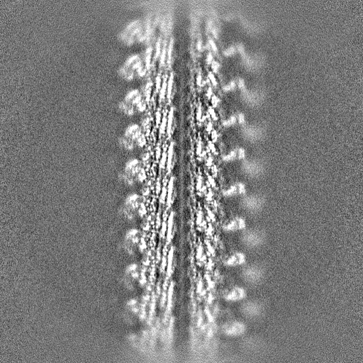
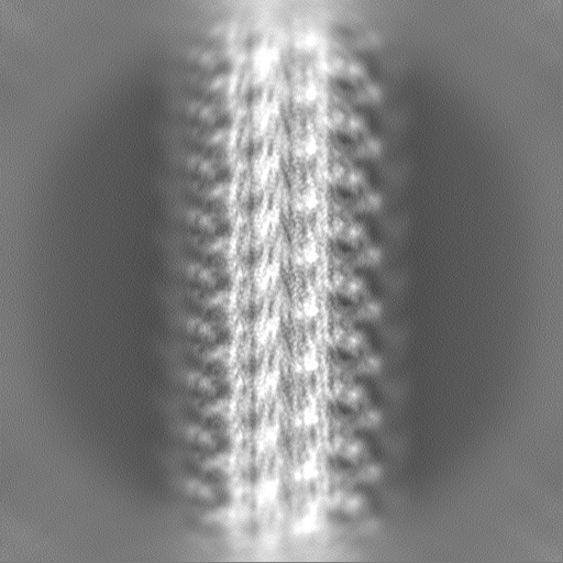
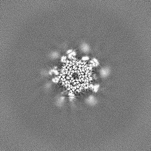
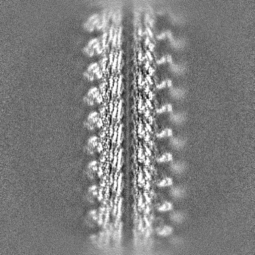
| Images |  emd_27059.png emd_27059.png | 92.3 KB | ||
| Others | emd_27059_half_map_1.map.gzemd_27059_half_map_2.map.gz | 476 MB 476 MB | ||
| Archive directory |  http://ftp.pdbj.org/pub/emdb/structures/EMD-27059ftp://ftp.pdbj.org/pub/emdb/structures/EMD-27059 http://ftp.pdbj.org/pub/emdb/structures/EMD-27059ftp://ftp.pdbj.org/pub/emdb/structures/EMD-27059 | HTTPS FTP |
-Related structure data


-Links
| EMDB pages | EMDB (EBI/PDBe) / EMDataResource |
|---|
-Map
| File | Download / File: emd_27059.map.gz / Format: CCP4 / Size: 512 MB / Type: IMAGE STORED AS FLOATING POINT NUMBER (4 BYTES) | ||||||||||||||||||||||||||||||||||||
|---|---|---|---|---|---|---|---|---|---|---|---|---|---|---|---|---|---|---|---|---|---|---|---|---|---|---|---|---|---|---|---|---|---|---|---|---|---|
| Annotation | Cryo-EM structure of the helical E. coli K12 flagellar filament core | ||||||||||||||||||||||||||||||||||||
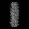
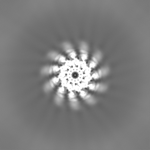
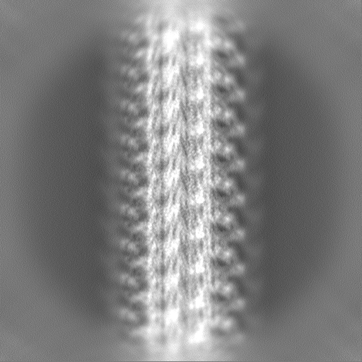
| Projections & slices | Image control
Images are generated by Spider. | ||||||||||||||||||||||||||||||||||||
| Voxel size | X=Y=Z: 1.08 Å | ||||||||||||||||||||||||||||||||||||
| Density |
| ||||||||||||||||||||||||||||||||||||
| Symmetry | Space group: 1 | ||||||||||||||||||||||||||||||||||||
| Details | EMDB XML:
|
 Z (Sec.)
 Y (Row.)
 X (Col.)


-Supplemental data
-Half map: Cryo-EM structure of the helical E. coli K12 flagellar filament core
| File | emd_27059_half_map_1.map | ||||||||||||
|---|---|---|---|---|---|---|---|---|---|---|---|---|---|
| Annotation | Cryo-EM structure of the helical E. coli K12 flagellar filament core | ||||||||||||
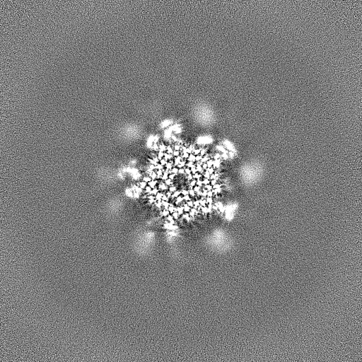
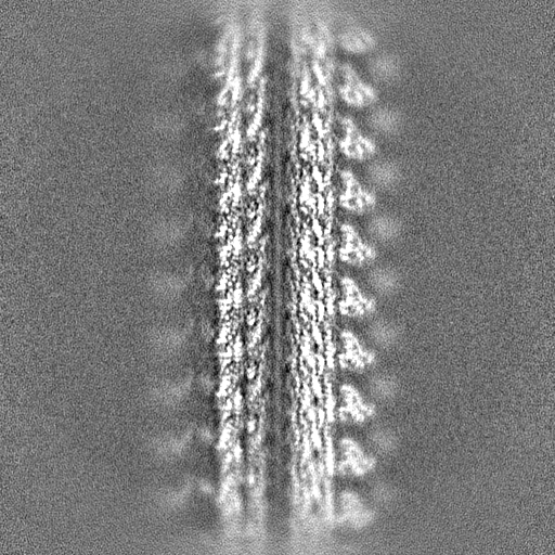
| Projections & Slices |
| ||||||||||||
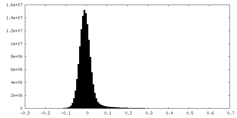
| Density Histograms |


-Half map: Cryo-EM structure of the helical E. coli K12 flagellar filament core
| File | emd_27059_half_map_2.map | ||||||||||||
|---|---|---|---|---|---|---|---|---|---|---|---|---|---|
| Annotation | Cryo-EM structure of the helical E. coli K12 flagellar filament core | ||||||||||||
| Projections & Slices |
| ||||||||||||
| Density Histograms |


- Sample components
-Entire : E. coli K12 flagellar filament core
| Entire | Name: E. coli K12 flagellar filament core |
|---|---|
| Components |
|
-Supramolecule #1: E. coli K12 flagellar filament core
| Supramolecule | Name: E. coli K12 flagellar filament core / type: complex / Chimera: Yes / ID: 1 / Parent: 0 / Macromolecule list: all |
|---|---|
| Source (natural) | Organism: |
-Macromolecule #1: Flagellin
| Macromolecule | Name: Flagellin / type: protein_or_peptide / ID: 1 / Enantiomer: LEVO |
|---|---|
| Sequence | String: MAQVINTNSL SLITQNNINK NQSALSSSIE RLSSGLRINS AKDDAAGQAI ANRFTSNIKG LTQAARNAN DGISVAQTTE GALSEINNNL QRVRELTVQA TTGTNSESDL SSIQDEIKSR L DEIDRVSG QTQFNGVNVL AKNGSMKIQV GANDNQTITI DLKQIDAKTL ...String: MAQVINTNSL SLITQNNINK NQSALSSSIE RLSSGLRINS AKDDAAGQAI ANRFTSNIKG LTQAARNAN DGISVAQTTE GALSEINNNL QRVRELTVQA TTGTNSESDL SSIQDEIKSR L DEIDRVSG QTQFNGVNVL AKNGSMKIQV GANDNQTITI DLKQIDAKTL GLDGFSVKNN DT VTTSAPV TAFGATTTNN IKLTGITLST EAATDTGGTN PASIEGVYTD NGNDYYAKIT GGD NDGKYY AVTVANDGTV TMATGATANA TVTDANTTKA TTITSGGTPV QIDNTAGSAT ANLG AVSLV KLQDSKGNDT DTYALKDTNG NLYAADVNET TGAVSVKTIT YTDSSGAASS PTAVK LGGD DGKTEVVDID GKTYDSADLN GGNLQTGLTA GGEALTAVAN GKTTDPLKAL DDAIAS VDK FRSSLGAVQN RLDSAVTNLN NTTTNLSEAQ SRIQDADYAT EVSNMSKAQI IQQAGNS VL AKANQVPQQV LSLLQG |
-Experimental details
-Structure determination
| Method | cryo EM |
|---|---|
 Processing Processing | helical reconstruction |
| Aggregation state | filament |
-Sample preparation
| Buffer | pH: 7.2 |
|---|---|
| Vitrification | Cryogen name: ETHANE |
- Electron microscopy
| Microscope | TFS KRIOS |
|---|---|
| Image recording | Film or detector model: GATAN K3 (6k x 4k) / Average electron dose: 50.0 e/Å2 |
| Electron beam | Acceleration voltage: 300 kV / Electron source:  FIELD EMISSION GUN FIELD EMISSION GUN |
| Electron optics | Illumination mode: FLOOD BEAM / Imaging mode: BRIGHT FIELD / Nominal defocus max: 2.2 µm / Nominal defocus min: 1.2 µm |
| Experimental equipment |  Model: Titan Krios / Image courtesy: FEI Company |
-Image processing
| Final reconstruction | Applied symmetry - Helical parameters - Δz: 4.832 Å Applied symmetry - Helical parameters - Δ&Phi: 65.402 ° Applied symmetry - Helical parameters - Axial symmetry: C1 (asymmetric) Resolution.type: BY AUTHOR / Resolution: 3.0 Å / Resolution method: FSC 0.143 CUT-OFF / Number images used: 520264 |
|---|---|
| Final angle assignment | Type: NOT APPLICABLE |
| FSC plot (resolution estimation) |  |
