 ムービー
ムービー コントローラー


+ データを開く

- 基本情報
基本情報
| 登録情報 |  | |||||||||
|---|---|---|---|---|---|---|---|---|---|---|
| タイトル | Rhoptry secretory apparatus of Plasmodium falciparum with an associated rhoptry tip from a fused rhoptry pair | |||||||||
 マップデータ マップデータ | Rhoptry secretory apparatus of Plasmodium falciparum with an associated rhoptry tip from a fused rhoptry pair | |||||||||
 試料 試料 |
| |||||||||
 キーワード キーワード | Exocytosis Membrane Parasite / CELL INVASION | |||||||||
| 生物種 |  | |||||||||
| 手法 | サブトモグラム平均法 / クライオ電子顕微鏡法 / 解像度: 50.0 Å | |||||||||
 データ登録者 データ登録者 | Martinez M / Chang Y-W | |||||||||
| 資金援助 |  米国, 1件 米国, 1件
| |||||||||
 引用 引用 | ジャーナル: Nat Microbiol / 年: 2022 タイトル: Rhoptry secretion system structure and priming in Plasmodium falciparum revealed using in situ cryo-electron tomography. 著者: Matthew Martinez / William David Chen / Marta Mendonça Cova / Petra Molnár / Shrawan Kumar Mageswaran / Amandine Guérin / Audrey R Odom John / Maryse Lebrun / Yi-Wei Chang /  要旨: Apicomplexan parasites secrete contents of the rhoptries, club-shaped organelles in the apical region, into host cells to permit their invasion and establishment of infection. The rhoptry secretory ...Apicomplexan parasites secrete contents of the rhoptries, club-shaped organelles in the apical region, into host cells to permit their invasion and establishment of infection. The rhoptry secretory apparatus (RSA), which is critical for rhoptry secretion, was recently discovered in Toxoplasma and Cryptosporidium. It is unknown whether a similar molecular machinery exists in the malaria parasite Plasmodium. In this study, we use in situ cryo-electron tomography to investigate the rhoptry secretion system in P. falciparum merozoites. We identify the presence of an RSA at the cell apex and a morphologically distinct apical vesicle docking the tips of the two rhoptries to the RSA. We also discover two additional rhoptry organizations that lack the apical vesicle. Using subtomogram averaging, we reveal different conformations of the RSA structure corresponding to different rhoptry organizations. Our results highlight previously unknown steps in the process of rhoptry secretion and indicate a regulatory role for the conserved apical vesicle in host invasion by apicomplexan parasites. | |||||||||
| 履歴 |
|
- 構造の表示
構造の表示
| 添付画像 |
|---|

- ダウンロードとリンク
-EMDBアーカイブ
| マップデータ | emd_26672.map.gz | 2.5 MB |  EMDBマップデータ形式 EMDBマップデータ形式 | |
|---|---|---|---|---|
| ヘッダ (付随情報) | emd-26672-v30.xmlemd-26672.xml | 18.5 KB 18.5 KB | 表示 表示 | EMDBヘッダ |
| FSC (解像度算出) | emd_26672_fsc.xml | 3.3 KB | 表示 | FSCデータファイル |
| 画像 | 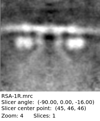 emd_26672.png emd_26672.png | 66.8 KB | ||
| マスクデータ | emd_26672_msk_1.map | 2.8 MB | マスクマップ | |
| Filedesc metadata | emd-26672.cif.gz | 5 KB | ||
| その他 | emd_26672_half_map_1.map.gzemd_26672_half_map_2.map.gz | 1.3 MB 1.3 MB | ||
| アーカイブディレクトリ |  http://ftp.pdbj.org/pub/emdb/structures/EMD-26672ftp://ftp.pdbj.org/pub/emdb/structures/EMD-26672 http://ftp.pdbj.org/pub/emdb/structures/EMD-26672ftp://ftp.pdbj.org/pub/emdb/structures/EMD-26672 | HTTPS FTP |
-検証レポート
| 文書・要旨 | emd_26672_validation.pdf.gz | 782 KB | 表示 | EMDB検証レポート |
|---|---|---|---|---|
| 文書・詳細版 | emd_26672_full_validation.pdf.gz | 781.6 KB | 表示 | |
| XML形式データ | emd_26672_validation.xml.gz | 8.9 KB | 表示 | |
| CIF形式データ | emd_26672_validation.cif.gz | 11.5 KB | 表示 | |
| アーカイブディレクトリ | https://ftp.pdbj.org/pub/emdb/validation_reports/EMD-26672ftp://ftp.pdbj.org/pub/emdb/validation_reports/EMD-26672 | HTTPS FTP |
-関連構造データ

-リンク
| EMDBのページ | EMDB (EBI/PDBe) / EMDataResource |
|---|
-マップ
| ファイル | ダウンロード / ファイル: emd_26672.map.gz / 形式: CCP4 / 大きさ: 2.8 MB / タイプ: IMAGE STORED AS FLOATING POINT NUMBER (4 BYTES) | ||||||||||||||||||||
|---|---|---|---|---|---|---|---|---|---|---|---|---|---|---|---|---|---|---|---|---|---|
| 注釈 | Rhoptry secretory apparatus of Plasmodium falciparum with an associated rhoptry tip from a fused rhoptry pair | ||||||||||||||||||||
| ボクセルのサイズ | X=Y=Z: 10.6 Å | ||||||||||||||||||||
| 密度 |
| ||||||||||||||||||||
| 対称性 | 空間群: 1 | ||||||||||||||||||||
| 詳細 | EMDB XML:
|
-添付データ
-マスク #1
| ファイル | emd_26672_msk_1.map | ||||||||||||
|---|---|---|---|---|---|---|---|---|---|---|---|---|---|
| 投影像・断面図 |
| ||||||||||||
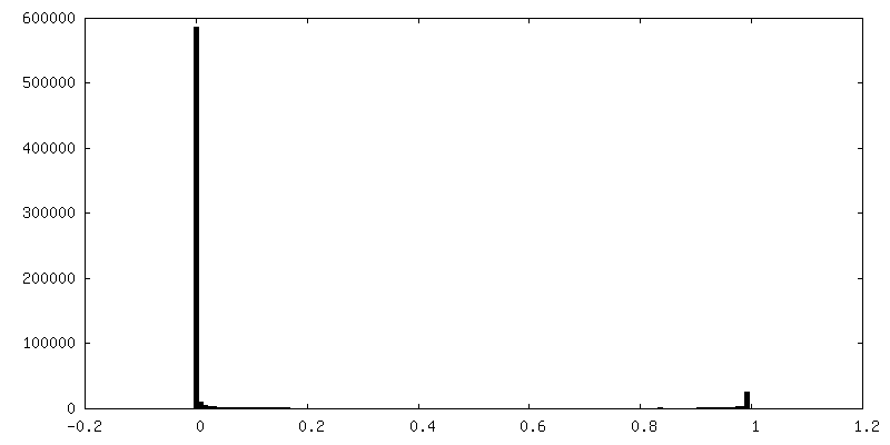
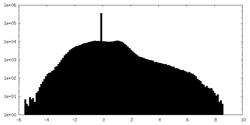
| 密度ヒストグラム |
 Z
Z Y
Y X
X


-ハーフマップ: Rhoptry secretory apparatus of Plasmodium falciparum with an...
| ファイル | emd_26672_half_map_1.map | ||||||||||||
|---|---|---|---|---|---|---|---|---|---|---|---|---|---|
| 注釈 | Rhoptry secretory apparatus of Plasmodium falciparum with an associated rhoptry tip from a fused rhoptry pair | ||||||||||||
| 投影像・断面図 |
| ||||||||||||
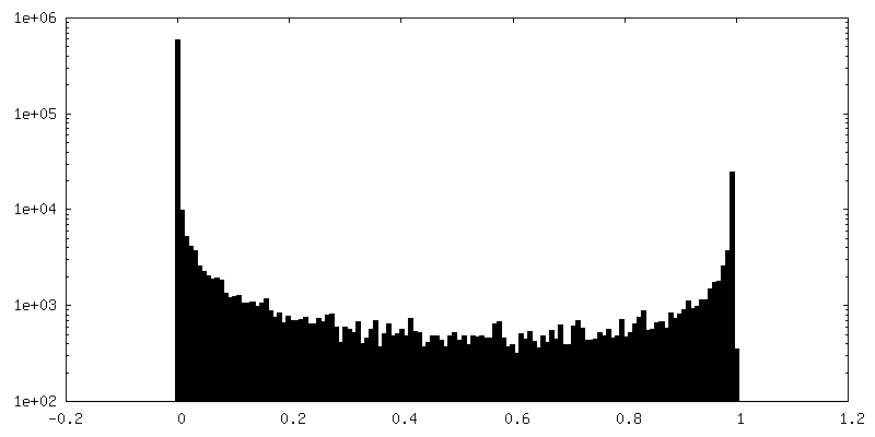
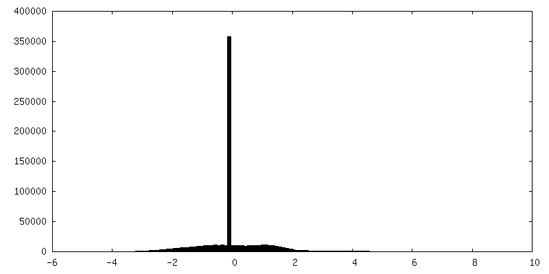
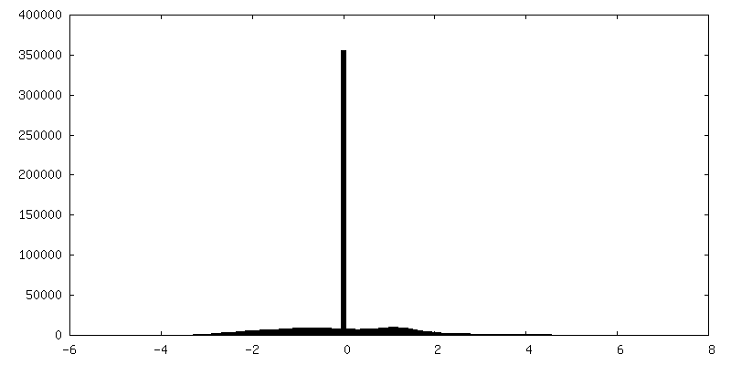
| 密度ヒストグラム |


-ハーフマップ: Rhoptry secretory apparatus of Plasmodium falciparum with an...
| ファイル | emd_26672_half_map_2.map | ||||||||||||
|---|---|---|---|---|---|---|---|---|---|---|---|---|---|
| 注釈 | Rhoptry secretory apparatus of Plasmodium falciparum with an associated rhoptry tip from a fused rhoptry pair | ||||||||||||
| 投影像・断面図 |
| ||||||||||||
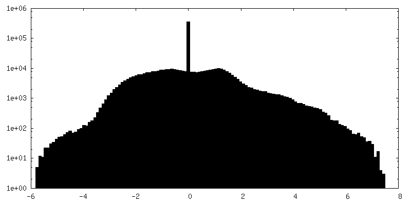
| 密度ヒストグラム |


- 試料の構成要素
-全体 : Plasmodium falciparum merozoite
| 全体 | 名称: Plasmodium falciparum merozoite |
|---|---|
| 要素 |
|
-超分子 #1: Plasmodium falciparum merozoite
| 超分子 | 名称: Plasmodium falciparum merozoite / タイプ: cell / ID: 1 / 親要素: 0 / 含まれる分子: #1-#2 |
|---|---|
| 由来(天然) | 生物種: |
-超分子 #2: Rhoptry secretory apparatus
| 超分子 | 名称: Rhoptry secretory apparatus / タイプ: complex / ID: 2 / 親要素: 1 / 含まれる分子: #1 詳細: 8-fold symmetric protein structure, anchored into the plasma membrane, which docks a fused rhoptry tip to the site of exocytosis at the plasma membrane |
|---|---|
| 由来(天然) | 生物種: Organelle: Apical complex / 細胞中の位置: Cytoplasm |
-超分子 #3: Rhoptry
| 超分子 | 名称: Rhoptry / タイプ: organelle_or_cellular_component / ID: 3 / 親要素: 1 / 含まれる分子: #2 詳細: The rhoptry tip from a fused rhoptry pair docked at the rhoptry secretory apparatus |
|---|
-実験情報
-構造解析
| 手法 | クライオ電子顕微鏡法 |
|---|---|
 解析 解析 | サブトモグラム平均法 |
| 試料の集合状態 | cell |
-試料調製
| 緩衝液 | pH: 7.4 詳細: RPMI medium modified with 27 mM NaHCO3, 11 mM glucose, 5 mM HEPES, 0.01 mM thymidine, 1 mM sodium pyruvate, 0.37 mM hypoxanthine, 10 ug/mL gentamicin, and 5 g/L Albumax |
|---|---|
| グリッド | モデル: Quantifoil R2/2 / 材質: COPPER / メッシュ: 200 / 支持フィルム - 材質: CARBON / 支持フィルム - トポロジー: HOLEY / 前処理 - タイプ: GLOW DISCHARGE |
| 凍結 | 凍結剤: ETHANE-PROPANE / チャンバー内湿度: 95 % / チャンバー内温度: 297 K / 装置: LEICA EM GP 詳細: Blot for 4 or 6 seconds from the front before plunging. |
| 詳細 | Mechanically isolated merozoites from a highly synchronous culture of Plasmodium falciparum schizonts |
- 電子顕微鏡法
| 顕微鏡 | FEI TITAN KRIOS |
|---|---|
| 特殊光学系 | 位相板: VOLTA PHASE PLATE / エネルギーフィルター - 名称: GIF Bioquantum / エネルギーフィルター - スリット幅: 20 eV |
| 撮影 | フィルム・検出器のモデル: GATAN K3 (6k x 4k) / 撮影したグリッド数: 9 / 平均露光時間: 0.45 sec. / 平均電子線量: 2.5 e/Å2 詳細: Images were collected in dose-fractionation mode with 0.1 second exposure per frame, for a total of 0.4 or 0.5 seconds per tilt image |
| 電子線 | 加速電圧: 300 kV / 電子線源:  FIELD EMISSION GUN FIELD EMISSION GUN |
| 電子光学系 | C2レンズ絞り径: 100.0 µm / 照射モード: FLOOD BEAM / 撮影モード: BRIGHT FIELD / 最大 デフォーカス(公称値): 4.0 µm / 最小 デフォーカス(公称値): 1.0 µm / 倍率(公称値): 33000 |
| 試料ステージ | 試料ホルダーモデル: FEI TITAN KRIOS AUTOGRID HOLDER ホルダー冷却材: NITROGEN |
| 実験機器 |  モデル: Titan Krios / 画像提供: FEI Company |
-画像解析
| 最終 再構成 | 想定した対称性 - 点群: C8 (8回回転対称) / アルゴリズム: BACK PROJECTION / 解像度のタイプ: BY AUTHOR / 解像度: 50.0 Å / 解像度の算出法: FSC 0.143 CUT-OFF 詳細: FSC was calculated using the "adaptive bandpass filter" function in Dynamo. FSC was calculated between the two half maps using a mask that covered the rhoptry secretory apparatus structure ...詳細: FSC was calculated using the "adaptive bandpass filter" function in Dynamo. FSC was calculated between the two half maps using a mask that covered the rhoptry secretory apparatus structure and the top of the associated rhoptry tip. 使用したサブトモグラム数: 36 | ||||||
|---|---|---|---|---|---|---|---|
| 抽出 | トモグラム数: 36 / 使用した粒子像数: 36 参照モデル: Average of manually preoriented particles with no search refinement 手法: Volumes picked manually ソフトウェア:
詳細: In IMOD, volumes were manually selected and preoriented | ||||||
| 最終 角度割当 | タイプ: ANGULAR RECONSTITUTION / ソフトウェア - 名称: Dynamo (ver. 1.1.509) | ||||||
| FSC曲線 (解像度の算出) | 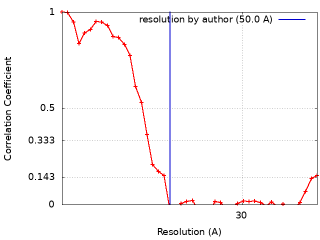 |
