 Movie
Movie Controller

[English] 日本語
 Yorodumi
- EMDB-26671: Rhoptry secretory apparatus of Plasmodium falciparum with an asso... -
+ Open data

- Basic information
| Entry |  | |||||||||
|---|---|---|---|---|---|---|---|---|---|---|
| Title | Rhoptry secretory apparatus of Plasmodium falciparum with an associated rhoptry tip from a non-fused rhoptry pair | |||||||||
 Map data Map data | Rhoptry secretory apparatus of Plasmodium falciparum with an associated rhoptry tip from a non-fused rhoptry pair | |||||||||
 Sample Sample |
| |||||||||
 Keywords Keywords | Exocytosis Membrane Parasite / CELL INVASION | |||||||||
| Biological species |  | |||||||||
| Method | subtomogram averaging / cryo EM / Resolution: 50.0 Å | |||||||||
 Authors Authors | Martinez M / Chang Y-W | |||||||||
| Funding support |  United States, 1 items United States, 1 items
| |||||||||
 Citation Citation | Journal: Nat Microbiol / Year: 2022 Title: Rhoptry secretion system structure and priming in Plasmodium falciparum revealed using in situ cryo-electron tomography. Authors: Matthew Martinez / William David Chen / Marta Mendonça Cova / Petra Molnár / Shrawan Kumar Mageswaran / Amandine Guérin / Audrey R Odom John / Maryse Lebrun / Yi-Wei Chang /  Abstract: Apicomplexan parasites secrete contents of the rhoptries, club-shaped organelles in the apical region, into host cells to permit their invasion and establishment of infection. The rhoptry secretory ...Apicomplexan parasites secrete contents of the rhoptries, club-shaped organelles in the apical region, into host cells to permit their invasion and establishment of infection. The rhoptry secretory apparatus (RSA), which is critical for rhoptry secretion, was recently discovered in Toxoplasma and Cryptosporidium. It is unknown whether a similar molecular machinery exists in the malaria parasite Plasmodium. In this study, we use in situ cryo-electron tomography to investigate the rhoptry secretion system in P. falciparum merozoites. We identify the presence of an RSA at the cell apex and a morphologically distinct apical vesicle docking the tips of the two rhoptries to the RSA. We also discover two additional rhoptry organizations that lack the apical vesicle. Using subtomogram averaging, we reveal different conformations of the RSA structure corresponding to different rhoptry organizations. Our results highlight previously unknown steps in the process of rhoptry secretion and indicate a regulatory role for the conserved apical vesicle in host invasion by apicomplexan parasites. | |||||||||
| History |
|
- Structure visualization
| Supplemental images |
|---|
- Downloads & links
-EMDB archive
| Map data | emd_26671.map.gz | 2.5 MB |  EMDB map data format EMDB map data format | |
|---|---|---|---|---|
| Header (meta data) | emd-26671-v30.xmlemd-26671.xml | 18.2 KB 18.2 KB | Display Display | EMDB header |
| FSC (resolution estimation) | emd_26671_fsc.xml | 3.3 KB | Display | FSC data file |
| Images |  emd_26671.png emd_26671.png | 62.1 KB | ||
| Masks | emd_26671_msk_1.map | 2.8 MB | Mask map | |
| Filedesc metadata | emd-26671.cif.gz | 5 KB | ||
| Others | emd_26671_half_map_1.map.gzemd_26671_half_map_2.map.gz | 1.3 MB 1.3 MB | ||
| Archive directory |  http://ftp.pdbj.org/pub/emdb/structures/EMD-26671ftp://ftp.pdbj.org/pub/emdb/structures/EMD-26671 http://ftp.pdbj.org/pub/emdb/structures/EMD-26671ftp://ftp.pdbj.org/pub/emdb/structures/EMD-26671 | HTTPS FTP |
-Related structure data


-Links
| EMDB pages | EMDB (EBI/PDBe) / EMDataResource |
|---|
-Map
| File | Download / File: emd_26671.map.gz / Format: CCP4 / Size: 2.8 MB / Type: IMAGE STORED AS FLOATING POINT NUMBER (4 BYTES) | ||||||||||||||||||||||||||||||||||||
|---|---|---|---|---|---|---|---|---|---|---|---|---|---|---|---|---|---|---|---|---|---|---|---|---|---|---|---|---|---|---|---|---|---|---|---|---|---|
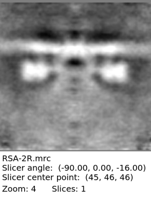
| Annotation | Rhoptry secretory apparatus of Plasmodium falciparum with an associated rhoptry tip from a non-fused rhoptry pair | ||||||||||||||||||||||||||||||||||||
| Projections & slices | Image control
Images are generated by Spider. | ||||||||||||||||||||||||||||||||||||
| Voxel size | X=Y=Z: 10.6 Å | ||||||||||||||||||||||||||||||||||||
| Density |
| ||||||||||||||||||||||||||||||||||||
| Symmetry | Space group: 1 | ||||||||||||||||||||||||||||||||||||
| Details | EMDB XML:
|
 Z (Sec.)
 Y (Row.)
 X (Col.)


-Supplemental data
-Mask #1
| File | emd_26671_msk_1.map | ||||||||||||
|---|---|---|---|---|---|---|---|---|---|---|---|---|---|
| Projections & Slices |
| ||||||||||||
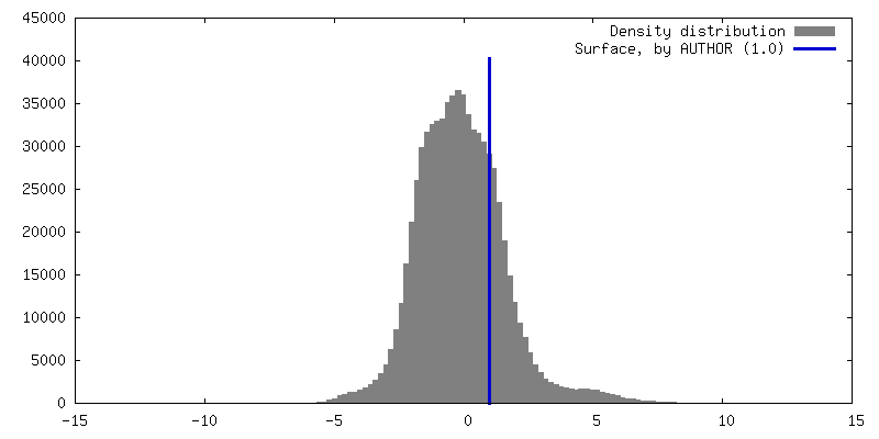
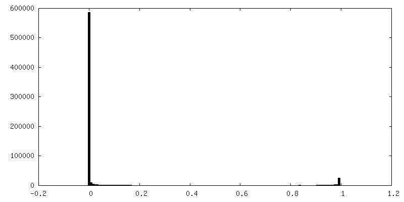
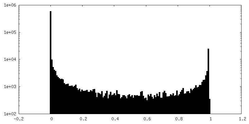
| Density Histograms |


-Half map: Rhoptry secretory apparatus of Plasmodium falciparum with an...
| File | emd_26671_half_map_1.map | ||||||||||||
|---|---|---|---|---|---|---|---|---|---|---|---|---|---|
| Annotation | Rhoptry secretory apparatus of Plasmodium falciparum with an associated rhoptry tip from a non-fused rhoptry pair | ||||||||||||
| Projections & Slices |
| ||||||||||||
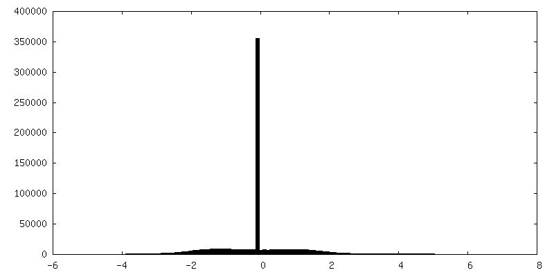
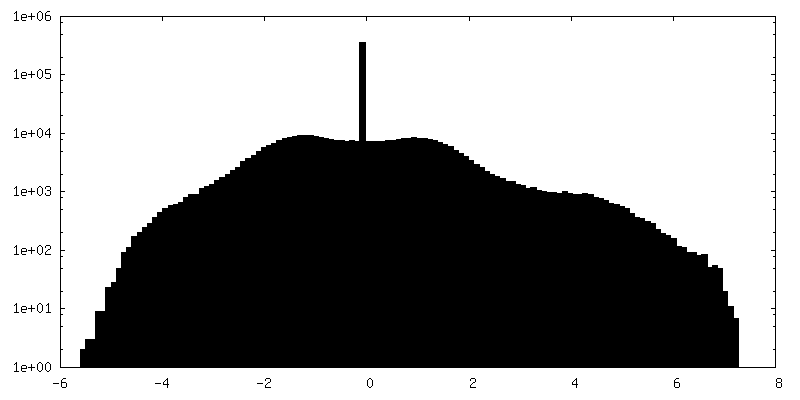
| Density Histograms |


-Half map: Rhoptry secretory apparatus of Plasmodium falciparum with an...
| File | emd_26671_half_map_2.map | ||||||||||||
|---|---|---|---|---|---|---|---|---|---|---|---|---|---|
| Annotation | Rhoptry secretory apparatus of Plasmodium falciparum with an associated rhoptry tip from a non-fused rhoptry pair | ||||||||||||
| Projections & Slices |
| ||||||||||||
| Density Histograms |


- Sample components
-Entire : Plasmodium falciparum merozoite
| Entire | Name: Plasmodium falciparum merozoite |
|---|---|
| Components |
|
-Supramolecule #1: Plasmodium falciparum merozoite
| Supramolecule | Name: Plasmodium falciparum merozoite / type: cell / ID: 1 / Parent: 0 / Macromolecule list: #1-#2 |
|---|---|
| Source (natural) | Organism: |
-Supramolecule #2: Rhoptry secretory apparatus
| Supramolecule | Name: Rhoptry secretory apparatus / type: complex / ID: 2 / Parent: 1 / Macromolecule list: #1 Details: 8-fold symmetric protein structure, anchored into the plasma membrane, which docks a rhoptry tip to the to the site of exocytosis at the plasma membrane |
|---|---|
| Source (natural) | Organism: |
-Supramolecule #3: Rhoptry tip
| Supramolecule | Name: Rhoptry tip / type: organelle_or_cellular_component / ID: 3 / Parent: 1 / Macromolecule list: #2 Details: One of two rhoptry tips that is docked at the rhoptry secretory apparatus. |
|---|
-Experimental details
-Structure determination
| Method | cryo EM |
|---|---|
 Processing Processing | subtomogram averaging |
| Aggregation state | cell |
-Sample preparation
| Buffer | pH: 7.4 Details: RPMI medium modified with 27 mM NaHCO3, 11 mM glucose, 5 mM HEPES, 0.01 mM thymidine, 1 mM sodium pyruvate, 0.37 mM hypoxanthine, 10 ug/mL gentamicin, and 5 g/L Albumax |
|---|---|
| Grid | Model: Quantifoil R2/2 / Material: COPPER / Mesh: 200 / Support film - Material: CARBON / Support film - topology: HOLEY / Pretreatment - Type: GLOW DISCHARGE |
| Vitrification | Cryogen name: ETHANE-PROPANE / Chamber humidity: 95 % / Chamber temperature: 297 K / Instrument: LEICA EM GP Details: Blot for 4 or 6 seconds from the front before plunging. |
| Details | Mechanically isolated merozoites from a highly synchronous culture of Plasmodium falciparum schizonts |
- Electron microscopy
| Microscope | FEI TITAN KRIOS |
|---|---|
| Specialist optics | Phase plate: VOLTA PHASE PLATE / Energy filter - Name: GIF Bioquantum / Energy filter - Slit width: 20 eV |
| Image recording | Film or detector model: GATAN K3 (6k x 4k) / Number grids imaged: 9 / Average exposure time: 0.45 sec. / Average electron dose: 2.5 e/Å2 Details: Images were collected in dose-fractionation mode with 0.1 second exposure per frame, for a total of 0.4 or 0.5 seconds per tilt image |
| Electron beam | Acceleration voltage: 300 kV / Electron source:  FIELD EMISSION GUN FIELD EMISSION GUN |
| Electron optics | C2 aperture diameter: 100.0 µm / Illumination mode: FLOOD BEAM / Imaging mode: BRIGHT FIELD / Nominal defocus max: 4.0 µm / Nominal defocus min: 1.0 µm / Nominal magnification: 33000 |
| Sample stage | Specimen holder model: FEI TITAN KRIOS AUTOGRID HOLDER / Cooling holder cryogen: NITROGEN |
| Experimental equipment |  Model: Titan Krios / Image courtesy: FEI Company |
-Image processing
| Final reconstruction | Applied symmetry - Point group: C8 (8 fold cyclic) / Algorithm: BACK PROJECTION / Resolution.type: BY AUTHOR / Resolution: 50.0 Å / Resolution method: FSC 0.143 CUT-OFF Details: FSC was calculated using the "adaptive bandpass filtering" function in Dynamo. The FSC was calculated using a mask that covers the rhoptry secretory apparatus structure and the top portion ...Details: FSC was calculated using the "adaptive bandpass filtering" function in Dynamo. The FSC was calculated using a mask that covers the rhoptry secretory apparatus structure and the top portion of the associated rhoptry tip. Number subtomograms used: 33 | ||||||
|---|---|---|---|---|---|---|---|
| Extraction | Number tomograms: 33 / Number images used: 33 Reference model: Average of manually preoriented particles with no search refinement Method: Volumes picked manually Software:
Details: In IMOD, volumes were manually selected and preoriented | ||||||
| Final angle assignment | Type: ANGULAR RECONSTITUTION / Software - Name: Dynamo (ver. 1.1.509) | ||||||
| FSC plot (resolution estimation) | 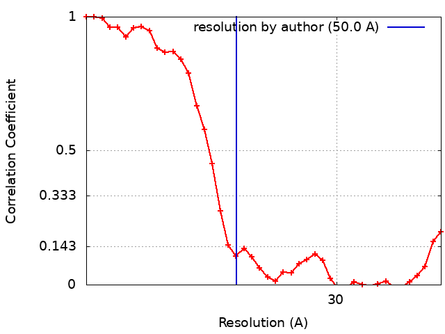 |
