ジャーナル: Nat Commun / 年: 2014 タイトル: The palindromic DNA-bound USP/EcR nuclear receptor adopts an asymmetric organization with allosteric domain positioning. 著者: Massimiliano Maletta / Igor Orlov / Pierre Roblin / Yannick Beck / Dino Moras / Isabelle M L Billas / Bruno P Klaholz / 要旨: Nuclear receptors (NRs) regulate gene expression through DNA- and ligand-binding and thus represent crucial therapeutic targets. The ultraspiracle protein/ecdysone receptor (USP/EcR) complex binds to ...Nuclear receptors (NRs) regulate gene expression through DNA- and ligand-binding and thus represent crucial therapeutic targets. The ultraspiracle protein/ecdysone receptor (USP/EcR) complex binds to half-sites with a one base pair spaced inverted repeat (IR1), a palindromic DNA response element (RE) reminiscent of IRs observed for vertebrate steroid hormone receptors. Here we present the cryo electron microscopy structure of the USP/EcR complex bound to an IR1 RE which provides the first description of a full IR-bound NR complex. The structure reveals that even though the DNA is almost symmetric, the complex adopts a highly asymmetric architecture in which the ligand-binding domains (LBDs) are positioned 5' off-centred. Additional interactions of the USP LBD with the 5'-flanking sequence trigger transcription activity as monitored by transfection assays. The comparison with DR-bound NR complexes suggests that DNA is the major allosteric driver in inversely positioning the LBDs, which serve as the main binding-site for transcriptional regulators.
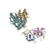
The domains (USP/EcR LBD and DBD heterodimers respectively, PDB IDs 1R1K and 2HAN) were separately fitted by manual docking using program Pymol.
精密化
空間: REAL / プロトコル: RIGID BODY FIT
得られたモデル
PDB-4umm: The Cryo-EM structure of the palindromic DNA-bound USP-EcR nuclear receptor reveals an asymmetric organization with allosteric domain positioning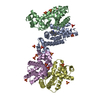
 ムービー
ムービー コントローラー


 データを開く
 基本情報
基本情報 マップデータ
 試料
試料 キーワード
キーワード 機能・相同性情報
 Heliothis virescens (蝶・蛾) / synthetic construct (人工物)
 データ登録者
 引用
引用
 構造の表示
構造の表示
 ダウンロードとリンク
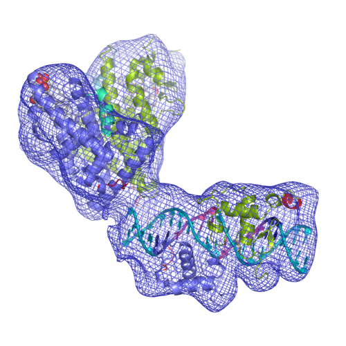 EMD-2631-USP-EcR-Image.png
 http://ftp.pdbj.org/pub/emdb/structures/EMD-2631


 Z (Sec.)
 Y (Row.)
 X (Col.)


 試料の構成要素
 解析
解析 電子顕微鏡法
 FIELD EMISSION GUN