 Movie
Movie Controller

[English] 日本語
 Yorodumi
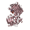
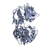
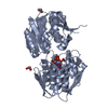
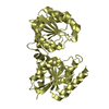
- PDB-1s7n: Ribosomal L7/L12 alpha-N-protein acetyltransferase in complex wit... -
+ Open data

- Basic information
| Entry | Database: PDB / ID: 1s7n | ||||||
|---|---|---|---|---|---|---|---|
| Title | Ribosomal L7/L12 alpha-N-protein acetyltransferase in complex with Coenzyme A (CoA free sulfhydryl) | ||||||
 Components Components | acetyl transferase | ||||||
 Keywords Keywords | TRANSFERASE / acetyltransferase / GNAT / alpha-N-protein acetyltransferase / Coenzyme A / L7/L12 | ||||||
| Function / homology |  Function and homology information Function and homology informationprotein N-terminal-serine acetyltransferase activity / protein-N-terminal-alanine acetyltransferase activity / Transferases; Acyltransferases; Transferring groups other than aminoacyl groups / cytoplasm Similarity search - Function | ||||||
| Biological species |  Salmonella typhimurium (bacteria) Salmonella typhimurium (bacteria) | ||||||
| Method |  X-RAY DIFFRACTION / MOLECULAR REPLACEMENT / Resolution: 2.1 Å X-RAY DIFFRACTION / MOLECULAR REPLACEMENT / Resolution: 2.1 Å | ||||||
 Authors Authors | Vetting, M.W. / de Carvalho, L.P. / Roderick, S.L. / Blanchard, J.S. | ||||||
 Citation Citation | Journal: J.Biol.Chem. / Year: 2005 Title: A novel dimeric structure of the RimL Nalpha-acetyltransferase from Salmonella typhimurium. Authors: Vetting, M.W. / de Carvalho, L.P. / Roderick, S.L. / Blanchard, J.S. | ||||||
| History |
|
- Structure visualization
| Structure viewer | Molecule: MolmilJmol/JSmol |
|---|
- Downloads & links
-Download
| PDBx/mmCIF format | 1s7n.cif.gz | 164 KB | Display | PDBx/mmCIF format |
|---|---|---|---|---|
| PDB format | pdb1s7n.ent.gz | 131 KB | Display | PDB format |
| PDBx/mmJSON format | 1s7n.json.gz | Tree view | PDBx/mmJSON format | |
| Others |  Other downloads Other downloads |
-Validation report
| Arichive directory | https://data.pdbj.org/pub/pdb/validation_reports/s7/1s7nftp://data.pdbj.org/pub/pdb/validation_reports/s7/1s7n | HTTPS FTP |
|---|
-Related structure data
| Related structure data | 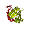 1s7fSC 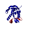 1s7kC 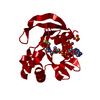 1s7lC S: Starting model for refinement C: citing same article ( |
|---|---|
| Similar structure data |


-Links
 PDBj
PDBj


- Assembly
| Deposited unit | 
| ||||||||
|---|---|---|---|---|---|---|---|---|---|
| 1 | 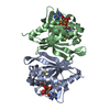
| ||||||||
| 2 | 
| ||||||||
| Unit cell |
| ||||||||
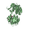
| Details | The biological assembly is a dimer. Monomers A/B and C/D make up the two physiological dimers in the asymmetric unit. |
-Components
| #1: Protein | Mass: 20912.771 Da / Num. of mol.: 4 Source method: isolated from a genetically manipulated source Source: (gene. exp.) Salmonella typhimurium (bacteria) / Strain: LT2 / Gene: RimL / Plasmid: pet28a+ / Production host: References: UniProt: Q8ZPC0, Transferases; Acyltransferases; Transferring groups other than aminoacyl groups #2: Chemical | ChemComp-COA /   Mass: 767.534 Da / Num. of mol.: 4 / Source method: obtained synthetically / Formula: C21H36N7O16P3S Mass: 767.534 Da / Num. of mol.: 4 / Source method: obtained synthetically / Formula: C21H36N7O16P3S#3: Water | ChemComp-HOH / |  Mass: 18.015 Da / Num. of mol.: 384 / Source method: isolated from a natural source / Formula: H2O Mass: 18.015 Da / Num. of mol.: 384 / Source method: isolated from a natural source / Formula: H2O |
|---|
-Experimental details
-Experiment
| Experiment | Method: X-RAY DIFFRACTION / Number of used crystals: 1 |
|---|
- Sample preparation
| Crystal | Density Matthews: 2.54 Å3/Da / Density % sol: 51.61 % |
|---|---|
| Crystal grow | Temperature: 293 K / Method: vapor diffusion under oil / pH: 8 Details: pentaerythritiol propoxylate 426, triethanolamine, KCl, ammonium sulfate, acetylCoenzyme A, TCEP, pH 8.0, vapor diffusion under oil, temperature 293K |
-Data collection
| Diffraction | Mean temperature: 77 K |
|---|---|
| Diffraction source | Source: ROTATING ANODE / Type: RIGAKU RU200 / Wavelength: 1.5418 Å |
| Detector | Type: RIGAKU RAXIS IV / Detector: IMAGE PLATE / Date: May 23, 2003 / Details: MSC Blue Confocal |
| Radiation | Monochromator: MSC Blue Confocal / Protocol: SINGLE WAVELENGTH / Monochromatic (M) / Laue (L): M / Scattering type: x-ray |
| Radiation wavelength | Wavelength: 1.5418 Å / Relative weight: 1 |
| Reflection | Resolution: 2.1→30 Å / Num. all: 45918 / Num. obs: 45918 / % possible obs: 93.7 % / Observed criterion σ(F): 0 / Observed criterion σ(I): 0 / Redundancy: 2.4 % / Biso Wilson estimate: 31.7 Å2 / Rsym value: 0.046 / Net I/σ(I): 16.9 |
| Reflection shell | Resolution: 2.1→2.18 Å / Redundancy: 2.1 % / Rsym value: 0.13 / % possible all: 86.6 |
- Processing
| Software |
| |||||||||||||||||||||||||
|---|---|---|---|---|---|---|---|---|---|---|---|---|---|---|---|---|---|---|---|---|---|---|---|---|---|---|
| Refinement | Method to determine structure: MOLECULAR REPLACEMENT Starting model: PDB ENTRY 1S7F Resolution: 2.1→30 Å / Cross valid method: THROUGHOUT / σ(F): 0 / σ(I): 0 / Stereochemistry target values: Engh & Huber
| |||||||||||||||||||||||||
| Refinement step | Cycle: LAST / Resolution: 2.1→30 Å
| |||||||||||||||||||||||||
| Refine LS restraints |
| |||||||||||||||||||||||||
| LS refinement shell | Resolution: 2.1→2.18 Å / Rfactor Rfree: 0.265 / Rfactor Rwork: 0.239 |
