 Movie
Movie Controller

[English] 日本語
 Yorodumi
- EMDB-26010: The symmetrized subpellicular microtubule map from intact Toxopla... -
+ Open data

- Basic information
| Entry | Database: EMDB / ID: EMD-26010 | ||||||||||||||||||
|---|---|---|---|---|---|---|---|---|---|---|---|---|---|---|---|---|---|---|---|
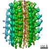
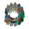
| Title | The symmetrized subpellicular microtubule map from intact Toxoplasma gondii cells | ||||||||||||||||||
 Map data Map data | |||||||||||||||||||
 Sample Sample |
| ||||||||||||||||||
 Keywords Keywords | parasites / Toxoplasma gondii / cytoskeleton / microtubules / CELL INVASION | ||||||||||||||||||
| Biological species |  | ||||||||||||||||||
| Method | subtomogram averaging / cryo EM / Resolution: 26.0 Å | ||||||||||||||||||
 Authors Authors | Sun SY / Chen M | ||||||||||||||||||
| Funding support |  United States, 5 items United States, 5 items
| ||||||||||||||||||
 Citation Citation | Journal: Proc Natl Acad Sci U S A / Year: 2022 Title: Cryo-ET of parasites gives subnanometer insight into tubulin-based structures. Authors: Stella Y Sun / Li-Av Segev-Zarko / Muyuan Chen / Grigore D Pintilie / Michael F Schmid / Steven J Ludtke / John C Boothroyd / Wah Chiu / Abstract: Tubulin is a conserved protein that polymerizes into different forms of filamentous structures in , an obligate intracellular parasite in the phylum Apicomplexa. Two key tubulin-containing ...Tubulin is a conserved protein that polymerizes into different forms of filamentous structures in , an obligate intracellular parasite in the phylum Apicomplexa. Two key tubulin-containing cytoskeletal components are subpellicular microtubules (SPMTs) and conoid fibrils (CFs). The SPMTs help maintain shape and gliding motility, while the CFs are implicated in invasion. Here, we use cryogenic electron tomography to determine the molecular structures of the SPMTs and CFs in vitrified intact and detergent-extracted parasites. Subvolume densities from detergent-extracted parasites yielded averaged density maps at subnanometer resolutions, and these were related back to their architecture in situ. An intralumenal spiral lines the interior of the 13-protofilament SPMTs, revealing a preferred orientation of these microtubules relative to the parasite's long axis. Each CF is composed of nine tubulin protofilaments that display a comma-shaped cross-section, plus additional associated components. Conoid protrusion, a crucial step in invasion, is associated with an altered pitch of each CF. The use of basic building blocks of protofilaments and different accessory proteins in one organism illustrates the versatility of tubulin to form two distinct types of assemblies, SPMTs and CFs. | ||||||||||||||||||
| History |
|
- Structure visualization
| Movie |
 Movie viewer Movie viewer |
|---|---|
| Structure viewer | EM map: SurfViewMolmilJmol/JSmol |
| Supplemental images |

- Downloads & links
-EMDB archive
| Map data | emd_26010.map.gz | 3.3 MB | EMDB map data format | |
|---|---|---|---|---|
| Header (meta data) | emd-26010-v30.xmlemd-26010.xml | 11.5 KB 11.5 KB | Display Display | EMDB header |
| Images | 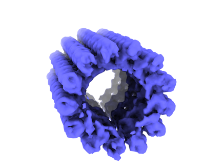 emd_26010.png emd_26010.png | 70 KB | ||
| Filedesc metadata | emd-26010.cif.gz | 4.3 KB | ||
| Archive directory |  http://ftp.pdbj.org/pub/emdb/structures/EMD-26010ftp://ftp.pdbj.org/pub/emdb/structures/EMD-26010 http://ftp.pdbj.org/pub/emdb/structures/EMD-26010ftp://ftp.pdbj.org/pub/emdb/structures/EMD-26010 | HTTPS FTP |
-Validation report
| Summary document | emd_26010_validation.pdf.gz | 399.9 KB | Display | EMDB validaton report |
|---|---|---|---|---|
| Full document | emd_26010_full_validation.pdf.gz | 399.5 KB | Display | |
| Data in XML | emd_26010_validation.xml.gz | 5.5 KB | Display | |
| Data in CIF | emd_26010_validation.cif.gz | 6.2 KB | Display | |
| Arichive directory | https://ftp.pdbj.org/pub/emdb/validation_reports/EMD-26010ftp://ftp.pdbj.org/pub/emdb/validation_reports/EMD-26010 | HTTPS FTP |
-Related structure data


-Links
| EMDB pages | EMDB (EBI/PDBe) / EMDataResource |
|---|
-Map
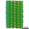
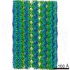
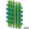
| File | Download / File: emd_26010.map.gz / Format: CCP4 / Size: 3.4 MB / Type: IMAGE STORED AS FLOATING POINT NUMBER (4 BYTES) | ||||||||||||||||||||||||||||||||||||||||||||||||||||||||||||||||||||
|---|---|---|---|---|---|---|---|---|---|---|---|---|---|---|---|---|---|---|---|---|---|---|---|---|---|---|---|---|---|---|---|---|---|---|---|---|---|---|---|---|---|---|---|---|---|---|---|---|---|---|---|---|---|---|---|---|---|---|---|---|---|---|---|---|---|---|---|---|---|
| Projections & slices | Image control
Images are generated by Spider. | ||||||||||||||||||||||||||||||||||||||||||||||||||||||||||||||||||||
| Voxel size | X=Y=Z: 7.086 Å | ||||||||||||||||||||||||||||||||||||||||||||||||||||||||||||||||||||
| Density |
| ||||||||||||||||||||||||||||||||||||||||||||||||||||||||||||||||||||
| Symmetry | Space group: 1 | ||||||||||||||||||||||||||||||||||||||||||||||||||||||||||||||||||||
| Details | EMDB XML:
CCP4 map header:
| ||||||||||||||||||||||||||||||||||||||||||||||||||||||||||||||||||||
 Z (Sec.)
 Y (Row.)
 X (Col.)


-Supplemental data
- Sample components
-Entire : In situ subpellicular microtubule of Toxoplasma gondii
| Entire | Name: In situ subpellicular microtubule of Toxoplasma gondii |
|---|---|
| Components |
|
-Supramolecule #1: In situ subpellicular microtubule of Toxoplasma gondii
| Supramolecule | Name: In situ subpellicular microtubule of Toxoplasma gondii type: organelle_or_cellular_component / ID: 1 / Parent: 0 |
|---|---|
| Source (natural) | Organism: |
-Experimental details
-Structure determination
| Method | cryo EM |
|---|---|
 Processing Processing | subtomogram averaging |
| Aggregation state | cell |
-Sample preparation
| Buffer | pH: 7.2 Details: Hanks balanced salts solution, supplemented (HBSS): 1 mM MgCl2, 1 mM CaCl2, 10 mM NaHCO3, 20 mM HEPES, pH 7.2 |
|---|---|
| Grid | Model: Homemade / Material: COPPER / Mesh: 200 / Support film - Material: CARBON / Support film - topology: LACEY / Pretreatment - Type: PLASMA CLEANING / Pretreatment - Time: 25 sec. |
| Vitrification | Cryogen name: ETHANE / Chamber humidity: 90 % / Chamber temperature: 295 K / Instrument: LEICA EM GP |
| Details | 3x10^6 cells/ml |
- Electron microscopy
| Microscope | FEI TALOS ARCTICA |
|---|---|
| Specialist optics | Phase plate: VOLTA PHASE PLATE / Energy filter - Name: GIF Bioquantum / Energy filter - Slit width: 20 eV |
| Image recording | Film or detector model: GATAN K2 SUMMIT (4k x 4k) / Detector mode: COUNTING / Average electron dose: 2.2 e/Å2 |
| Electron beam | Acceleration voltage: 200 kV / Electron source:  FIELD EMISSION GUN FIELD EMISSION GUN |
| Electron optics | C2 aperture diameter: 100.0 µm / Illumination mode: FLOOD BEAM / Imaging mode: BRIGHT FIELD / Cs: 2.7 mm / Nominal defocus max: 1.0 µm / Nominal defocus min: 1.0 µm / Nominal magnification: 39000 |
| Sample stage | Specimen holder model: FEI TITAN KRIOS AUTOGRID HOLDER / Cooling holder cryogen: NITROGEN |
| Experimental equipment |  Model: Talos Arctica / Image courtesy: FEI Company |
-Image processing
| Final reconstruction | Applied symmetry - Helical parameters - Δz: 80.0 Å Applied symmetry - Helical parameters - Δ&Phi: 27.7 ° Applied symmetry - Helical parameters - Axial symmetry: C13 (13 fold cyclic) Resolution.type: BY AUTHOR / Resolution: 26.0 Å / Resolution method: FSC 0.143 CUT-OFF / Software - Name: EMAN2 / Number subtomograms used: 1994 |
|---|---|
| Extraction | Number tomograms: 10 / Number images used: 1994 / Software - Name: EMAN2 |
| Final angle assignment | Type: OTHER |
