Movie
Movie Controller

[English] 日本語
 Yorodumi
- EMDB-24831: CryoEM structure of Methylocystis sp. str. Rockwell pMMO in a POP... -
+ Open data

- Basic information
| Entry |  | |||||||||
|---|---|---|---|---|---|---|---|---|---|---|
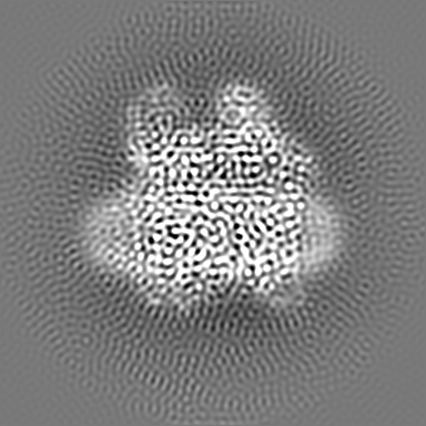
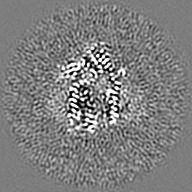
| Title | CryoEM structure of Methylocystis sp. str. Rockwell pMMO in a POPC nanodisc at 2.42 Angstrom resolution | |||||||||
 Map data Map data | CryoEM structure of Methylocystis species strain Rockwell pMMO in a POPC nanodisc at 2.42 angstrom resolution | |||||||||
 Sample Sample |
| |||||||||
 Keywords Keywords | Complex / OXIDOREDUCTASE | |||||||||
| Biological species |  Methylocystis sp. ATCC 49242 (bacteria) Methylocystis sp. ATCC 49242 (bacteria) | |||||||||
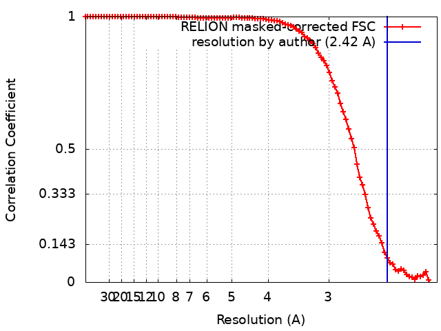
| Method | single particle reconstruction / cryo EM / Resolution: 2.42 Å | |||||||||
 Authors Authors | Koo CW / Rosenzweig AC | |||||||||
| Funding support |  United States, 1 items United States, 1 items
| |||||||||
 Citation Citation | Journal: Science / Year: 2022 Title: Recovery of particulate methane monooxygenase structure and activity in a lipid bilayer. Authors: Christopher W Koo / Frank J Tucci / Yuan He / Amy C Rosenzweig / Abstract: Bacterial methane oxidation using the enzyme particulate methane monooxygenase (pMMO) contributes to the removal of environmental methane, a potent greenhouse gas. Crystal structures determined using ...Bacterial methane oxidation using the enzyme particulate methane monooxygenase (pMMO) contributes to the removal of environmental methane, a potent greenhouse gas. Crystal structures determined using inactive, detergent-solubilized pMMO lack several conserved regions neighboring the proposed active site. We show that reconstituting pMMO in nanodiscs with lipids extracted from the native organism restores methane oxidation activity. Multiple nanodisc-embedded pMMO structures determined by cryo-electron microscopy to 2.14- to 2.46-angstrom resolution reveal the structure of pMMO in a lipid environment. The resulting model includes stabilizing lipids, regions of the PmoA and PmoC subunits not observed in prior structures, and a previously undetected copper-binding site in the PmoC subunit with an adjacent hydrophobic cavity. These structures provide a revised framework for understanding and engineering pMMO function. | |||||||||
| History |
|
- Structure visualization
| Supplemental images |
|---|

- Downloads & links
-EMDB archive
| Map data | emd_24831.map.gz | 201 MB |  EMDB map data format EMDB map data format | |
|---|---|---|---|---|
| Header (meta data) | emd-24831-v30.xmlemd-24831.xml | 16.9 KB 16.9 KB | Display Display | EMDB header |
| FSC (resolution estimation) | emd_24831_fsc.xml | 9.1 KB | Display | FSC data file |
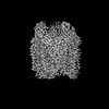
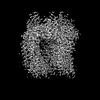
| Images | 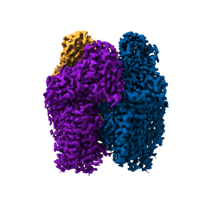 emd_24831.png emd_24831.png | 116.4 KB | ||
| Filedesc metadata | emd-24831.cif.gz | 5.9 KB | ||
| Others | emd_24831_additional_1.map.gz | 23.2 MB | ||
| Archive directory |  http://ftp.pdbj.org/pub/emdb/structures/EMD-24831ftp://ftp.pdbj.org/pub/emdb/structures/EMD-24831 http://ftp.pdbj.org/pub/emdb/structures/EMD-24831ftp://ftp.pdbj.org/pub/emdb/structures/EMD-24831 | HTTPS FTP |
-Related structure data
| Related structure data |  7s4mMC  7s4hC  7s4iC  7s4jC  7s4kC  7s4lC  7t4oC  7t4pC C: citing same article ( M: atomic model generated by this map |
|---|


-Links
| EMDB pages | EMDB (EBI/PDBe) / EMDataResource |
|---|
-Map
| File | Download / File: emd_24831.map.gz / Format: CCP4 / Size: 216 MB / Type: IMAGE STORED AS FLOATING POINT NUMBER (4 BYTES) | ||||||||||||||||||||||||||||||||||||
|---|---|---|---|---|---|---|---|---|---|---|---|---|---|---|---|---|---|---|---|---|---|---|---|---|---|---|---|---|---|---|---|---|---|---|---|---|---|
| Annotation | CryoEM structure of Methylocystis species strain Rockwell pMMO in a POPC nanodisc at 2.42 angstrom resolution | ||||||||||||||||||||||||||||||||||||
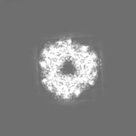
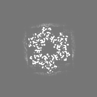
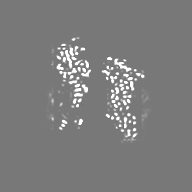
| Projections & slices | Image control
Images are generated by Spider. | ||||||||||||||||||||||||||||||||||||
| Voxel size | X=Y=Z: 0.52 Å | ||||||||||||||||||||||||||||||||||||
| Density |
| ||||||||||||||||||||||||||||||||||||
| Symmetry | Space group: 1 | ||||||||||||||||||||||||||||||||||||
| Details | EMDB XML:
|
 Z (Sec.)
 X (Row.)
 Y (Col.)


-Supplemental data
-Additional map: CryoEM structure of Methylocystis species strain Rockwell pMMO...
| File | emd_24831_additional_1.map | ||||||||||||
|---|---|---|---|---|---|---|---|---|---|---|---|---|---|
| Annotation | CryoEM structure of Methylocystis species strain Rockwell pMMO in a POPC nanodisc at 2.42 angstrom resolution processed by deepEMhancer | ||||||||||||
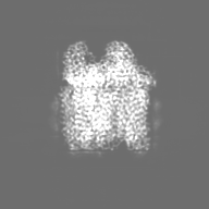
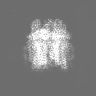
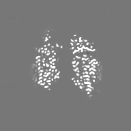
| Projections & Slices |
| ||||||||||||
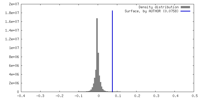
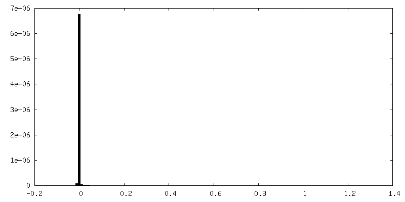
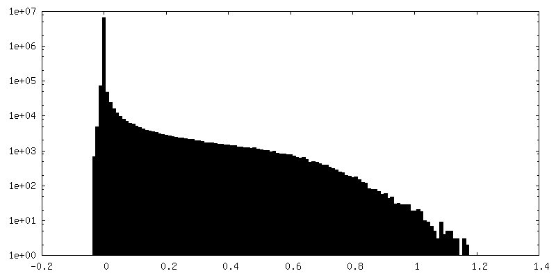
| Density Histograms |
- Sample components
+Entire : pMMO complex in a native lipid nanodisc
+Supramolecule #1: pMMO complex in a native lipid nanodisc
+Macromolecule #1: Particulate methane monooxygenase alpha subunit
+Macromolecule #2: Particulate methane monooxygenase beta subunit
+Macromolecule #3: Ammonia monooxygenase/methane monooxygenase, subunit C family protein
+Macromolecule #4: Unidentified Helix
+Macromolecule #5: COPPER (II) ION

+Macromolecule #6: 1,2-dihexanoyl-sn-glycero-3-phosphocholine
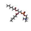
+Macromolecule #7: DECANE

+Macromolecule #8: DODECANE

+Macromolecule #9: water

-Experimental details
-Structure determination
| Method | cryo EM |
|---|---|
 Processing Processing | single particle reconstruction |
| Aggregation state | particle |
-Sample preparation
| Buffer | pH: 7.3 |
|---|---|
| Vitrification | Cryogen name: ETHANE |
- Electron microscopy
| Microscope | FEI TITAN KRIOS |
|---|---|
| Image recording | Film or detector model: GATAN K3 (6k x 4k) / Average electron dose: 1.02 e/Å2 |
| Electron beam | Acceleration voltage: 300 kV / Electron source:  FIELD EMISSION GUN FIELD EMISSION GUN |
| Electron optics | Illumination mode: FLOOD BEAM / Imaging mode: BRIGHT FIELD |
| Experimental equipment |  Model: Titan Krios / Image courtesy: FEI Company |