 ムービー
ムービー コントローラー

[日本語] English
 万見
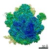
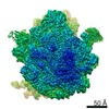
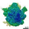
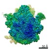
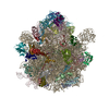
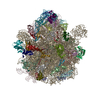
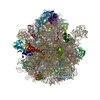
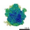
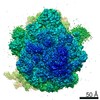
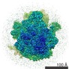
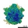
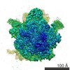
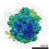
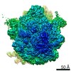
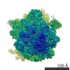
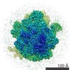
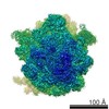
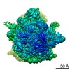
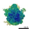
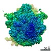
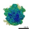
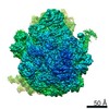
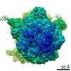
万見- EMDB-24800: wild-type Escherichia coli stalled ribosome with antibiotic linezolid -
+ データを開く

- 基本情報
基本情報
| 登録情報 | データベース: EMDB / ID: EMD-24800 | ||||||||||||||||||||||||||||||||||||
|---|---|---|---|---|---|---|---|---|---|---|---|---|---|---|---|---|---|---|---|---|---|---|---|---|---|---|---|---|---|---|---|---|---|---|---|---|---|
| タイトル | wild-type Escherichia coli stalled ribosome with antibiotic linezolid | ||||||||||||||||||||||||||||||||||||
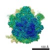
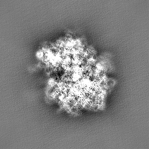
 マップデータ マップデータ | final unsharpened map used for model building | ||||||||||||||||||||||||||||||||||||
 試料 試料 |
| ||||||||||||||||||||||||||||||||||||
 キーワード キーワード | Escherichia coli stalled ribosome / oxazolidinone / linezolid / RIBOSOME-ANTIBIOTIC complex | ||||||||||||||||||||||||||||||||||||
| 機能・相同性 |  機能・相同性情報 機能・相同性情報positive regulation of ribosome biogenesis / DnaA-L2 complex / negative regulation of DNA-templated DNA replication initiation / cytosolic ribosome assembly / assembly of large subunit precursor of preribosome / regulation of cell growth / mRNA 5'-UTR binding / large ribosomal subunit / ribosomal small subunit assembly / transferase activity ...positive regulation of ribosome biogenesis / DnaA-L2 complex / negative regulation of DNA-templated DNA replication initiation / cytosolic ribosome assembly / assembly of large subunit precursor of preribosome / regulation of cell growth / mRNA 5'-UTR binding / large ribosomal subunit / ribosomal small subunit assembly / transferase activity / ribosome binding / 5S rRNA binding / small ribosomal subunit / ribosomal large subunit assembly / small ribosomal subunit rRNA binding / cytosolic small ribosomal subunit / cytosolic large ribosomal subunit / cytoplasmic translation / tRNA binding / negative regulation of translation / rRNA binding / structural constituent of ribosome / ribosome / translation / ribonucleoprotein complex / mRNA binding / RNA binding / zinc ion binding / membrane / metal ion binding / cytoplasm / cytosol 類似検索 - 分子機能 | ||||||||||||||||||||||||||||||||||||
| 生物種 |  | ||||||||||||||||||||||||||||||||||||
| 手法 | 単粒子再構成法 / クライオ電子顕微鏡法 / 解像度: 2.48 Å | ||||||||||||||||||||||||||||||||||||
 データ登録者 データ登録者 | Young ID / Stojkovic V / Tsai K / Lee DJ | ||||||||||||||||||||||||||||||||||||
| 資金援助 | 11件
| ||||||||||||||||||||||||||||||||||||
 引用 引用 | ジャーナル: Protein Sci / 年: 2021 タイトル: UCSF ChimeraX: Structure visualization for researchers, educators, and developers. 著者: Eric F Pettersen / Thomas D Goddard / Conrad C Huang / Elaine C Meng / Gregory S Couch / Tristan I Croll / John H Morris / Thomas E Ferrin /   要旨: UCSF ChimeraX is the next-generation interactive visualization program from the Resource for Biocomputing, Visualization, and Informatics (RBVI), following UCSF Chimera. ChimeraX brings (a) ...UCSF ChimeraX is the next-generation interactive visualization program from the Resource for Biocomputing, Visualization, and Informatics (RBVI), following UCSF Chimera. ChimeraX brings (a) significant performance and graphics enhancements; (b) new implementations of Chimera's most highly used tools, many with further improvements; (c) several entirely new analysis features; (d) support for new areas such as virtual reality, light-sheet microscopy, and medical imaging data; (e) major ease-of-use advances, including toolbars with icons to perform actions with a single click, basic "undo" capabilities, and more logical and consistent commands; and (f) an app store for researchers to contribute new tools. ChimeraX includes full user documentation and is free for noncommercial use, with downloads available for Windows, Linux, and macOS from https://www.rbvi.ucsf.edu/chimerax. | ||||||||||||||||||||||||||||||||||||
| 履歴 |
|
- 構造の表示
構造の表示
| ムービー |
ムービービューア |
|---|---|
| 構造ビューア | EMマップ: SurfViewMolmilJmol/JSmol |
| 添付画像 |

- ダウンロードとリンク
-EMDBアーカイブ
| マップデータ | emd_24800.map.gz | 474.3 MB | EMDBマップデータ形式 | |
|---|---|---|---|---|
| ヘッダ (付随情報) | emd-24800-v30.xmlemd-24800.xml | 76.3 KB 76.3 KB | 表示 表示 | EMDBヘッダ |
| FSC (解像度算出) | emd_24800_fsc.xml | 23.1 KB | 表示 | FSCデータファイル |
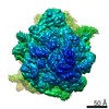
| 画像 |  emd_24800.png emd_24800.png | 155.8 KB | ||
| Filedesc metadata | emd-24800.cif.gz | 15.4 KB | ||
| アーカイブディレクトリ |  http://ftp.pdbj.org/pub/emdb/structures/EMD-24800ftp://ftp.pdbj.org/pub/emdb/structures/EMD-24800 http://ftp.pdbj.org/pub/emdb/structures/EMD-24800ftp://ftp.pdbj.org/pub/emdb/structures/EMD-24800 | HTTPS FTP |
-関連構造データ


-リンク
| EMDBのページ | EMDB (EBI/PDBe) / EMDataResource |
|---|---|
| 「今月の分子」の関連する項目 |


-マップ
| ファイル | ダウンロード / ファイル: emd_24800.map.gz / 形式: CCP4 / 大きさ: 512 MB / タイプ: IMAGE STORED AS FLOATING POINT NUMBER (4 BYTES) | ||||||||||||||||||||||||||||||||||||||||||||||||||||||||||||
|---|---|---|---|---|---|---|---|---|---|---|---|---|---|---|---|---|---|---|---|---|---|---|---|---|---|---|---|---|---|---|---|---|---|---|---|---|---|---|---|---|---|---|---|---|---|---|---|---|---|---|---|---|---|---|---|---|---|---|---|---|---|
| 注釈 | final unsharpened map used for model building | ||||||||||||||||||||||||||||||||||||||||||||||||||||||||||||
| 投影像・断面図 | 画像のコントロール
画像は Spider により作成 | ||||||||||||||||||||||||||||||||||||||||||||||||||||||||||||
| ボクセルのサイズ | X=Y=Z: 0.8125 Å | ||||||||||||||||||||||||||||||||||||||||||||||||||||||||||||
| 密度 |
| ||||||||||||||||||||||||||||||||||||||||||||||||||||||||||||
| 対称性 | 空間群: 1 | ||||||||||||||||||||||||||||||||||||||||||||||||||||||||||||
| 詳細 | EMDB XML:
CCP4マップ ヘッダ情報:
| ||||||||||||||||||||||||||||||||||||||||||||||||||||||||||||
 Z (Sec.)
 Y (Row.)
 X (Col.)


-添付データ
- 試料の構成要素
+全体 : wild-type Escherichia coli stalled ribosome with antibiotic linezolid
+超分子 #1: wild-type Escherichia coli stalled ribosome with antibiotic linezolid
+分子 #1: 30S ribosomal protein S19
+分子 #2: 30S ribosomal protein S20
+分子 #3: 30S ribosomal protein S21
+分子 #7: 30S ribosomal protein S2
+分子 #8: 30S ribosomal protein S3
+分子 #9: 30S ribosomal protein S4
+分子 #10: 30S ribosomal protein S5
+分子 #11: 30S ribosomal protein S6
+分子 #14: 50S ribosomal protein L2
+分子 #15: 50S ribosomal protein L3
+分子 #16: 50S ribosomal protein L4
+分子 #17: 50S ribosomal protein L5
+分子 #18: 50S ribosomal protein L6
+分子 #19: 50S ribosomal protein L9
+分子 #20: 50S ribosomal protein L31
+分子 #21: 50S ribosomal protein L13
+分子 #22: 50S ribosomal protein L14
+分子 #23: 50S ribosomal protein L15
+分子 #24: 50S ribosomal protein L16
+分子 #25: 50S ribosomal protein L17
+分子 #26: 50S ribosomal protein L18
+分子 #27: 50S ribosomal protein L19
+分子 #28: 50S ribosomal protein L20
+分子 #29: 50S ribosomal protein L21
+分子 #30: 50S ribosomal protein L22
+分子 #31: 50S ribosomal protein L23
+分子 #32: 50S ribosomal protein L24
+分子 #33: 50S ribosomal protein L25
+分子 #34: 50S ribosomal protein L27
+分子 #35: 50S ribosomal protein L28
+分子 #36: 50S ribosomal protein L29
+分子 #37: 50S ribosomal protein L30
+分子 #38: 50S ribosomal protein L32
+分子 #39: 50S ribosomal protein L33
+分子 #40: 50S ribosomal protein L34
+分子 #41: 50S ribosomal protein L35
+分子 #42: 50S ribosomal protein L36
+分子 #43: 30S ribosomal protein S7
+分子 #44: 30S ribosomal protein S8
+分子 #45: 30S ribosomal protein S9
+分子 #46: 30S ribosomal protein S10
+分子 #47: 30S ribosomal protein S11
+分子 #48: nascent peptide chain
+分子 #49: 30S ribosomal protein S12
+分子 #50: 30S ribosomal protein S13
+分子 #51: 30S ribosomal protein S14
+分子 #52: 30S ribosomal protein S15
+分子 #53: 30S ribosomal protein S16
+分子 #54: 30S ribosomal protein S17
+分子 #55: 30S ribosomal protein S18
+分子 #4: mRNA
+分子 #5: tRNA(PHE)
+分子 #6: 16S rRNA
+分子 #12: 23S rRNA
+分子 #13: 5S rRNA
+分子 #56: MAGNESIUM ION
+分子 #57: N-{[(5S)-3-(3-fluoro-4-morpholin-4-ylphenyl)-2-oxo-1,3-oxazolidin...

+分子 #58: ZINC ION
+分子 #59: water

-実験情報
-構造解析
| 手法 | クライオ電子顕微鏡法 |
|---|---|
 解析 解析 | 単粒子再構成法 |
| 試料の集合状態 | particle |
-試料調製
| 緩衝液 | pH: 7.5 |
|---|---|
| グリッド | モデル: Quantifoil R1.2/1.3 / 材質: COPPER / メッシュ: 300 / 支持フィルム - 材質: CARBON / 支持フィルム - トポロジー: CONTINUOUS / 支持フィルム - Film thickness: 2 / 前処理 - タイプ: GLOW DISCHARGE / 前処理 - 時間: 30 sec. / 詳細: 15 mA |
| 凍結 | 凍結剤: ETHANE / チャンバー内湿度: 95 % / チャンバー内温度: 283.2 K / 装置: FEI VITROBOT MARK IV |
- 電子顕微鏡法
| 顕微鏡 | FEI TITAN KRIOS |
|---|---|
| 撮影 | フィルム・検出器のモデル: GATAN K3 (6k x 4k) / 撮影したグリッド数: 1 / 実像数: 3644 / 平均電子線量: 67.8 e/Å2 |
| 電子線 | 加速電圧: 300 kV / 電子線源:  FIELD EMISSION GUN FIELD EMISSION GUN |
| 電子光学系 | 照射モード: FLOOD BEAM / 撮影モード: BRIGHT FIELD |
| 試料ステージ | 試料ホルダーモデル: FEI TITAN KRIOS AUTOGRID HOLDER ホルダー冷却材: NITROGEN |
| 実験機器 |  モデル: Titan Krios / 画像提供: FEI Company |
+画像解析

-原子モデル構築 1
| 精密化 | 空間: REAL / プロトコル: FLEXIBLE FIT |
|---|---|
| 得られたモデル | 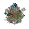 PDB-7s1g: |
