National Institutes of Health/National Institute of General Medical Sciences (NIH/NIGMS)
GM124348
United States
National Institutes of Health/National Institute of General Medical Sciences (NIH/NIGMS)
GM084210
United States
German Research Foundation (DFG)
AR1164/1-1
United States
American Heart Association
POST34030308
United States
Howard Hughes Medical Institute (HHMI)
United States
Citation
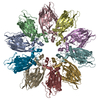
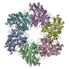
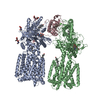
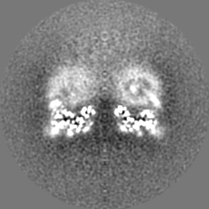
Journal: Nat Struct Mol Biol / Year: 2022 Title: Seipin forms a flexible cage at lipid droplet formation sites. Authors: Henning Arlt / Xuewu Sui / Brayden Folger / Carson Adams / Xiao Chen / Roman Remme / Fred A Hamprecht / Frank DiMaio / Maofu Liao / Joel M Goodman / Robert V Farese / Tobias C Walther / Abstract: Lipid droplets (LDs) form in the endoplasmic reticulum by phase separation of neutral lipids. This process is facilitated by the seipin protein complex, which consists of a ring of seipin monomers, ...Lipid droplets (LDs) form in the endoplasmic reticulum by phase separation of neutral lipids. This process is facilitated by the seipin protein complex, which consists of a ring of seipin monomers, with a yet unclear function. Here, we report a structure of S. cerevisiae seipin based on cryogenic-electron microscopy and structural modeling data. Seipin forms a decameric, cage-like structure with the lumenal domains forming a stable ring at the cage floor and transmembrane segments forming the cage sides and top. The transmembrane segments interact with adjacent monomers in two distinct, alternating conformations. These conformations result from changes in switch regions, located between the lumenal domains and the transmembrane segments, that are required for seipin function. Our data indicate a model for LD formation in which a closed seipin cage enables triacylglycerol phase separation and subsequently switches to an open conformation to allow LD growth and budding.
History
Deposition
Aug 11, 2021
-
Header (metadata) release
Feb 9, 2022
-
Map release
Feb 9, 2022
-
Update
Jun 5, 2024
-
Current status
Jun 5, 2024
Processing site: RCSB / Status: Released
-
Structure visualization
Movie
Surface view with section colored by density value
Name: Seipin; Sei1; Fld1 / type: complex / ID: 1 / Parent: 0 / Macromolecule list: all Details: Seipin complex; 10 subunits of monomers (chain A-J) in two alternating conformations
Source (natural)
Organism: Saccharomyces cerevisiae (brewer's yeast) / Strain: HAY60 / Organelle: ER / Location in cell: ER-LD junction
Molecular weight
Theoretical: 66 KDa
-
Supramolecule #2: Seipin dimer
Supramolecule
Name: Seipin dimer / type: complex / ID: 2 / Parent: 1 / Macromolecule list: all Details: Dimer of seipin monomers in transmembrane segment conformation A and B (chain A and B). That was used to build the oligomer consisting of 5 dimers (10 monomers).
Source (natural)
Organism: Saccharomyces cerevisiae (brewer's yeast) / Strain: HAY60 / Organelle: ER / Location in cell: ER-LD junction
-
Macromolecule #1: Seipin
Macromolecule
Name: Seipin / type: protein_or_peptide / ID: 1 / Number of copies: 10 / Enantiomer: LEVO
Cryogen name: ETHANE / Chamber humidity: 100 % / Chamber temperature: 277 K / Instrument: FEI VITROBOT MARK IV
Details
This sample was monodisperse.
-
Electron microscopy
Microscope
FEI TITAN KRIOS
Image recording
Film or detector model: GATAN K3 (6k x 4k) / Number grids imaged: 1 / Number real images: 12655 / Average exposure time: 1.9 sec. / Average electron dose: 54.59 e/Å2
Electron beam
Acceleration voltage: 300 kV / Electron source: OTHER
In the structure databanks used in Yorodumi, some data are registered as the other names, "COVID-19 virus" and "2019-nCoV". Here are the details of the virus and the list of structure data.
Jan 31, 2019. EMDB accession codes are about to change! (news from PDBe EMDB page)
EMDB accession codes are about to change! (news from PDBe EMDB page)
The allocation of 4 digits for EMDB accession codes will soon come to an end. Whilst these codes will remain in use, new EMDB accession codes will include an additional digit and will expand incrementally as the available range of codes is exhausted. The current 4-digit format prefixed with “EMD-” (i.e. EMD-XXXX) will advance to a 5-digit format (i.e. EMD-XXXXX), and so on. It is currently estimated that the 4-digit codes will be depleted around Spring 2019, at which point the 5-digit format will come into force.
The EM Navigator/Yorodumi systems omit the EMD- prefix.
Related info.:Q: What is EMD? / ID/Accession-code notation in Yorodumi/EM Navigator
Yorodumi is a browser for structure data from EMDB, PDB, SASBDB, etc.
This page is also the successor to EM Navigator detail page, and also detail information page/front-end page for Omokage search.
The word "yorodu" (or yorozu) is an old Japanese word meaning "ten thousand". "mi" (miru) is to see.
Related info.:EMDB / PDB / SASBDB / Comparison of 3 databanks / Yorodumi Search / Aug 31, 2016. New EM Navigator & Yorodumi / Yorodumi Papers / Jmol/JSmol / Function and homology information / Changes in new EM Navigator and Yorodumi
 Movie
Movie Controller


 Open data
 Basic information
 Map data
 Sample
 Keywords
 Function and homology information
 Authors
 United States, 5 items
  Citation
 Structure visualization
 UCSF Chimera
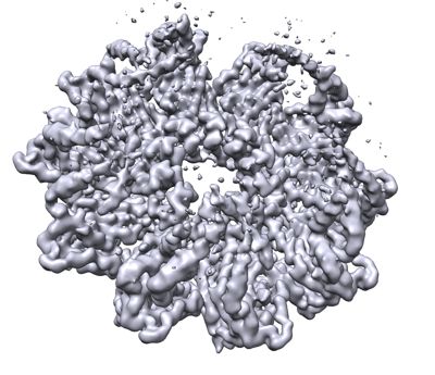
 Downloads & links
 emd_24674.png
 http://ftp.pdbj.org/pub/emdb/structures/EMD-24674


 Z (Sec.)
 Y (Row.)
 X (Col.)


 Sample components
 Processing
 Electron microscopy
